Preparations, Electrode placement – ZOLL R Series Monitor Defibrillator Rev J User Manual
Page 452
C
HAPTER
9
ECG M
ONITORING
9–2
www.zoll.com
9650-0912-01 Rev. K
Caution
ECG electrodes embedded in OneStep Pacing and Complete resuscitation pads produce
non-standard ECG monitoring lead vectors, designated P1, P2 and P3. While ECG signals
acquired from these leads are appropriate for ECG rhythm assessment and determining
electrical capture during pacing, they should not be used for ECG morphological
evaluations. Attach conventional ECG electrodes for diagnostic purposes.
Preparations
Proper application and placement of electrodes is essential for high quality ECG monitoring.
Good contact between the electrode and skin minimizes motion artifact and signal interference.
Remove all clothing covering the patient’s chest. Dry chest if necessary. If the patient has
excessive chest hair, clip or shave it to ensure proper adhesion of the electrodes.
Electrode Placement
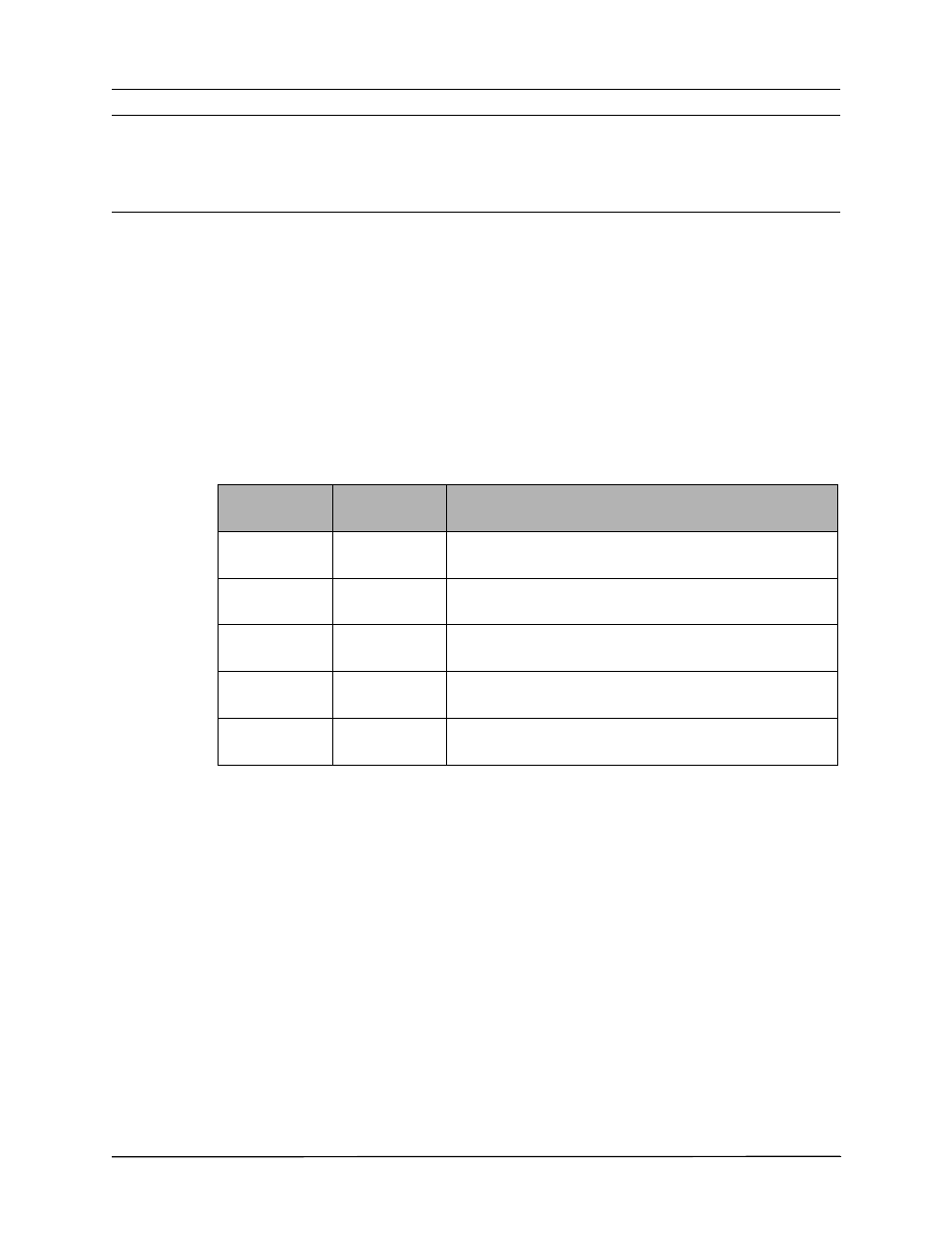
Depending upon local usage, the ECG leads are marked either RA, LA, LL, RL, and V or R, L,
F, N and C. The following table shows the markings and color codes for the different lead sets.
* Not used for 3-lead monitoring
IEC Color
Coding
AHA Color
Coding
Placement of Electrodes
R/Red Electrode RA/White
Electrode
Place near patient’s right mid-clavicular line, directly below
clavicle.
L/Yellow
Electrode
LA/Black
Electrode
Place near patient’s left mid-clavicular line, directly below
clavicle.
F/Green
Electrode
LL/Red
Electrode
Place between 6th and 7th intercostal space on patient’s left
mid-clavicular line.
N/Black*
Electrode
RL/Green*
Electrode
Place between 6th and 7th intercostal space on patient’s right
mid-clavicular line.
C/White*
Electrode
V/Brown*
Electrode
Single movable chest electrode.