Merit Medical HeartSpan Fixed Curve Braided Transseptal Sheath User Manual
Page 3
3
3. While the sheath and dilator hubs are separated,
slowly remove the guidewire from the dilator. Remove
all air from the dilator by slowly aspirating blood. After
making certain no air is in the dilator, flush the dilator.
4. Completely flush the transseptal needle.
5. Introduce the needle into the dilator hub. Carefully
advance the curved section of the needle into the
dilator, being certain not to restrict the movement
of the needle.
6. Withdraw the sheath approximately one centimeter
while maintaining the dilator position. Re-attach the
dilator and sheath hubs.
7. While maintaining the sheath’s position, slowly
advance the curved section of the needle until it is
about to protrude from the dilator tip.
8. Monitor the right atrial pressure by connecting the
needle hub to pressure monitoring equipment.
Good right atrial pressure should be observed before
proceeding.
9. Position the needle and sheath set in the right atrium.
Verify position using fluoroscopy.
10. Position the unit (dilator and needle point) against
the atrial septum in the region of the fossa ovale by
gradually rotating the needle posteriorly and toward
the left scapula during withdraw. Use continual
pressure monitoring and repeated anterior-posterior
and lateral visualization of the tip under fluoroscopy
during all positioning procedures.
11. After confirming the position of the dilator tip and
needle point against the atrial septum, advance
the needle and complete the transseptal puncture.
Successful needle entry into the left atrium is
confirmed by pressure monitoring and a sudden
reduction in resistance. It is critical that acceptable
left atrial pressure monitoring immediately after
the needle penetration is felt through the interatrial
septum. Do not advance the dilator if acceptable
pressure is not seen. Disconnect the pressure
monitoring line from the needle. This will show
the location of the needle. Reconnect the pressure
monitoring line to the needle.
12. Advance the dilator with the needle in place through
the septum. Acceptable left atrial pressure should
be continually observed. A sequential increase in
resistance to movement followed by a sharp decrease
in resistance will indicate location of the dilator in
the left atrium.
13. Withdraw the needle point even with the dilator
tip. The dilator with the needle point within should
be freely located in the left atrium. Verify with
fluoroscopy.
14. Advance the sheath slowly over the dilator-needle
combination until it is in the left atrium. A slow
rotating motion of the sheath as firm pressure is
applied will aid in this procedure. The sheath will be in
position when a sharp reduction of resistance is felt.
15. Advance the sheath approximately 2 cm into the left
atrium while maintaining the dilator needle position.
16. Disconnect the pressure monitoring line from the
needle.
17. Slowly remove the needle from the dilator.
18. Slowly remove the dilator from the sheath.
19. Attach the sheath sideport to the monitoring line.
Gently aspirate blood through the side arm for
sampling and to be sure the sheath is clear of air.
Caution: Remove the dilator slowly to reduce the
possibility of creating a vacuum in the sheath. Blood
should aspirate freely through the sideport. If not,
withdraw the sheath 0.5-1.0 cm (sheath tip may be
resting against the wall of the atrium or a pulmonary
vein). Note: Do not apply strong vacuum.
20. In order to maintain the location of the sheath in the
left atrium, monitor the location of the radiopaque tip
marker frequently under the fluoroscopy.
21. Introduce the properly prepared catheter through
the hemostatic valve into the left atrium. Improved
catheter manipulation may be obtained by
withdrawing the sheath into the right atrium. The
sheath should be returned to the left atrium over the
catheter prior to removing the catheter. Location of
sheath can be confirmed by comparison to position
established in step 20.
22. After removal of the sheath, use standard technique to
achieve hemostasis.
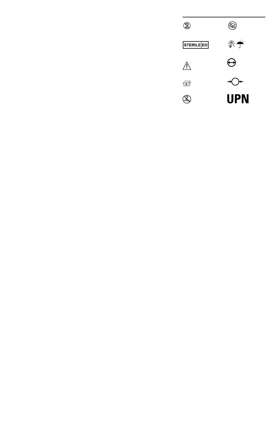
Single Use Only /
Do Not Reuse
Contents non-pyrogenic and
sterilized using ethylene
oxide
Caution, read intructions for
use prior to use
Contents (Numeral
represents quantity of units
inside)
Do Not Resterilize
Do not use if package is
opened or damaged
Store in a cool, dark and
dry place
Inner Diameter
Outer Diameter
Product Number