4 semi-dry transfer using the trans-blot sd cell, 3 total protein blot stains, 4 semi-dry transfer using the trans-blot – Bio-Rad Criterion™ TBE-Urea Precast Gels User Manual
Page 42: Sd cell
36
Technical Support: 1-800-4BIORAD • 1-800-424-6723 • www.bio-rad.com
Method
Sensitivity
Protein
Load
(μg/Band)
Advantages
Disadvantages
Imaging
SYPRO Ruby
protein blot stain
2–8 ng
~0.2
Compatible with
mass spectrometry,
Edman-based
sequencing,
and standard
immunological
procedures
Multi-step protocol;
requires UV, LED,
or laser imaging for
maximum sensitivity
Fluorescence
visualization
with UV, LED
epi-illumination or
laser scanning
Colloidal gold stain
1 ng
~0.1
Highly sensitive,
single-step protocol
Incompatible with nylon
membranes
Photography with
epi-illumination
or reflectance
densitometry
Anionic dyes
(amido black,
Coomassie R-250,
Ponceau S, Fast
Green FCF)
100–1,000 ng
~5.0
Inexpensive, rapid
Low sensitivity
Table 13.1. Total protein blot stains.
13.2.4 Semi-Dry Transfer Using the Trans-Blot
®
SD Cell
1. Equilibrate the gels and membranes (for example, in transfer buffer; see Appendix B for buffer
recipes) for 20 min prior to blot assembly.
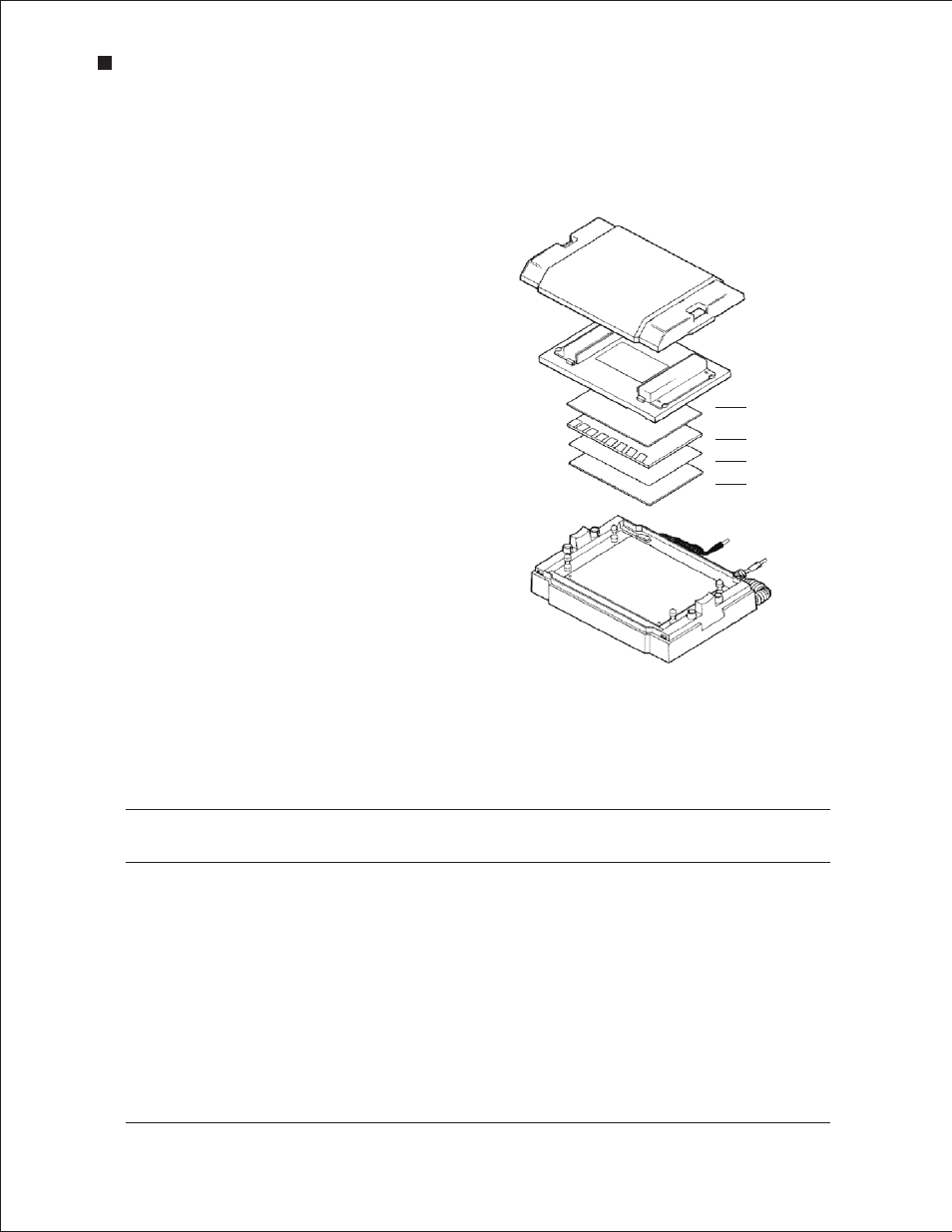
2. Assemble the blot for transfer using the Trans-Blot SD semi-dry transfer system.
(–)
(+)
Membrane
Gel
Filter paper
Filter paper
3. Connect the Trans-Blot SD cell to a PowerPac HC
power supply and begin transfer at 10–15 V.
For most proteins transferred from Criterion precast
gels, optimum transfer efficiency is obtained in
30 min; smaller proteins (<30 kD) may transfer more
quickly, while proteins >150 kD may show increased
transfer efficiencies at up to 60 min. Run times longer
than 60 min are NOT recommended for semi-dry
transfers.
Refer to the Trans-Blot SD Instruction Manual (bulletin
1703940) or the Protein Blotting Guide (bulletin 2895)
for additional information.
13.3 Total Protein Blot Stains
Total protein staining of a membrane provides
an image of the complete protein pattern, which
is required for the full characterization of specific
antigens detected in complex protein mixtures.
Gels shrink during staining, so comparison of an
immunologically probed membrane to a stained gel is
not practical. Instead, the exact location of a specifc
antigen is determined by comparing two blotted
membranes: one that has been probed with an
antibody and the other stained for total protein.
Assembly of the gel blot sandwich with the
Trans-Blot SD cell .
Criterion Precast Gels
- Criterion™ TBE Precast Gels Criterion™ Cell Criterion Precast Gels Criterion Dodeca Cell 2-D Electrophoresis Workflow Criterion™ IEF Precast Gels Criterion™ Zymogram Precast Gels Criterion™ XT Tris-Acetate Precast Gels Criterion™ Tris-Tricine Precast Gels Criterion™ XT Bis-Tris Precast Gels Criterion Stain Free™ Tris-HCl Gels Criterion™ Tris-HCl Precast Gels Criterion™ TGX Stain-Free™ Precast Gels Criterion™ TGX™ Precast Gels