Bio-Rad CHEF-DR II System User Manual
Page 31
6. The DNA is transferred from the back of the gel (the side opposite the wells) onto the
membrane because irregularities in the surface of the gel frequently occur during solidi-
fication of these high percentage gels (1%). These surface artifacts will interfere with the
transfer of the DNAs from the gel. Transfer from the other side of the gel insures smooth
surface contact between the gel and the membrane.
7. It is essential to neutralize the membrane after transfer to prevent changing the pH of the
hybridization buffer during hybridization.
8. It is not absolutely necessary to bake nylon membranes after alkaline transfer since the
DNA should be fixed onto the membrane by NaOH.
9. To monitor the efficiency of the transfer, stain the gel in neutralization buffer for 30 min-
utes with 1 µg/ml EtBr. Take a photograph of the post-transferred gel, and compare with
the original picture.
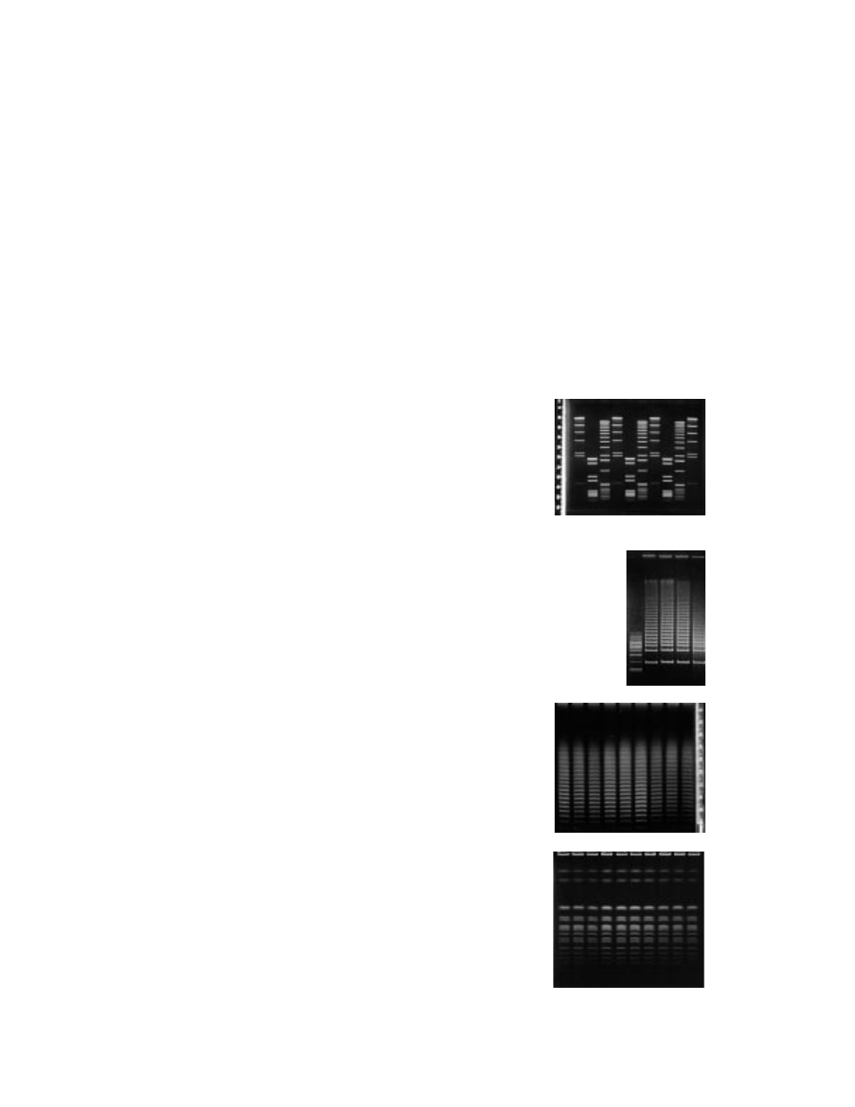
5.5 Separations of DNA Size Standards
1.
Restriction fragments
Size Range:
0.2-23 kb
Agarose:
1.0% Molecular Biology Certified
Buffer:
0.5x TBE
Temperature:
14 °C
Switch Time:
0.1 second
Run Time:
4 hours
Voltage Gradient: 6 V/cm
2.
5 kb Ladder
Size Range:
5-75 kb
Agarose:
1.0% Molecular Biology Certified
Buffer:
0.5x TBE
Temperature:
14 °C
Switch Time:
1-6 seconds
Run Time:
11 hours
Voltage Gradient: 6 V/cm
3.
Lambda Ladder
Size Range:
50-1000 kb
Agarose:
1.0% Molecular Biology Certified
Buffer:
0.5x TBE
Temperature:
14 °C
Switch Time:
50-90 seconds
Run Time:
22 hours
Voltage Gradient: 6 V/cm
4.
Saccharomyces cerevisiae
Size Range:
240-2200 kb
Agarose:
1.0% Pulsed Field Certified
Buffer:
0.5x TBE
Temperature:
14 °C
Switch Time:
60-120 seconds
Run Time:
24 hours
Voltage Gradient: 6 V/cm
28