Bio-Rad Helios® Gene Gun System User Manual
Page 37
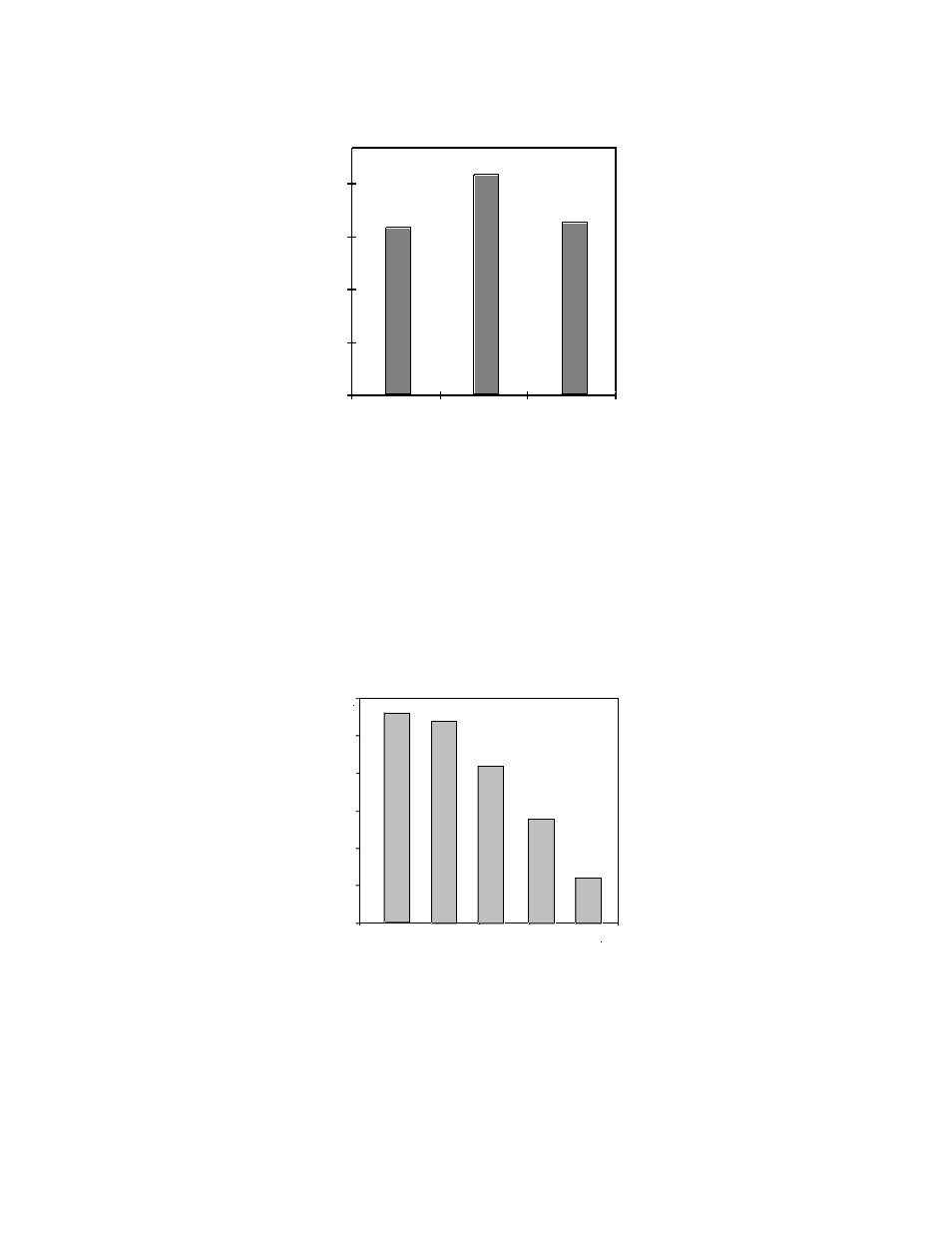
Fig. 20. Luciferase expression in mouse skin transfected
in vivo using the Helios Gene Gun.
Skin homogenates were assayed 24 hours post-transfection.
As illustrated in Figure 21, the optimum MLQ is determined by adjusting the concentration
of the DNA/microcarrier suspension loaded into the Gold-Coat tubing. For suspension cells,
higher MLQs (0.5–1.0 mg gold/cartridge) are often needed due to the high cell concentrations in
the aqueous cell smear. Adherent cell monolayers and intact tissues may require reduced MLQs
(0.06–0.25 mg gold/cartridge) to minimize tissue damage while maximizing transfection.
Fig. 21. Effect of the amount of accelerated gold particles on
in vitro expression levels.
Suspension
cultures of CHO cells were bombarded as described in Section 6.2 using the Accell Gene Gun. After 18 hours,
the CHO cells that had reattached to the tissue culture plate were lysed and assayed for luciferase expression
(Thompson
et al. 1993).
33
0.0
0.5
1.0
1.5
2.0
2.5
3.0
mg gold per cartridge
Luciferase relative light units x 10
-8
1.0
0.5
0.25
0.12
0.06
-6
100
200
400
Helium pressure (psi)
80
60
40
20
0
hGH activity (ng/ml)
Luciferase activity (relative light units x 10
-8
)