PASCO CI-6539A EKG SENSOR User Manual
Page 7
012–06852A
EKG Sensor
3
simultaneously. This process causes a small time delay and so there
is a short pause after the atria contract before the ventricles contract.
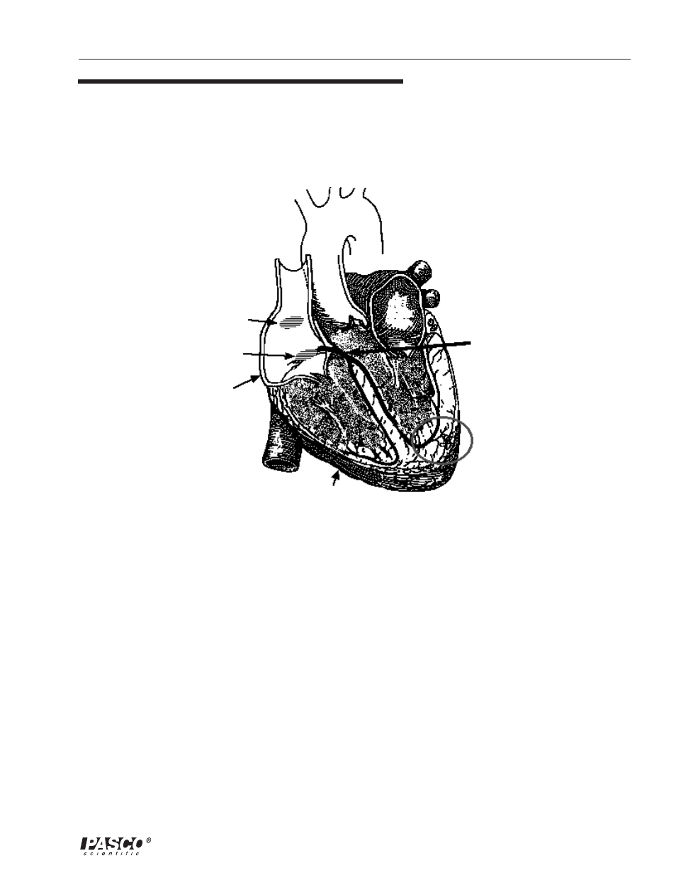
Because the cells of the heart muscle are interconnected, this wave of
depolarization, contraction and repolarization spreads across all the
connected muscle of the heart. See Figure 2.
Bundle of His
(Atrioventricular Bundle)
Purkinje Fibers
(Conduction Myofibers)
Right Ventricle
Right Atrium
Atrioventricular (AV)
Node
Sinoatrial (SA)
Node
Figure 2
Cross section of human heart
When a portion of the heart is polarized and the adjacent portion is
depolarized this creates an electrical current that moves through the
body. This current is greatest when one half of the connected portion
of the heart is polarized and the adjacent half is not polarized. The
current decreases when the ratio of polarized tissue to non-polarized
tissue is less than one-to-one. The changes in these currents can be
measured, amplified, and plotted over time. The EKG represents the
summation of all the actions potentials from the heart as detected on
the surface of the body and does not measure the mechanical
contractions of the heart directly.
The two atria contract due to the pacemaker and force blood into the
two ventricles. Shortly after this contraction the two ventricles
contract due to the signal conducted to them from the atria. The blood
leaves the two ventricles through pulmonary and aortic arteries. The
heart muscle cells recover their polarity and in another second the
cycle starts again.
➤
➤
➤
➤
➤ Note: An excellent text about the
electrocardiogram and other
phenomena of bioelectricity is Physics
with Health Science Applications by
Paul Peter Urone, ©1986, John Wiley
& Sons, Inc., New York.