Ac b – Bio-Rad Fluorescent Protein Stains User Manual
Page 11
9
R
2
= 0.9943
0
500
1000
1500
2000
2500
0
1
2
3
4
5
fl
u
o
rescen
ce
R
2
= 0.9947
0
5000
10000
15000
20000
25000
0
10
20
30
40
50
60
70
ng
β-galactosidase
fl
u
o
rescen
ce
R
2
= 0.9960
0
50000
100000
150000
200000
250000
300000
0
200
400
600
800
1000
1200
ng
β-galactosidase
fl
u
o
rescen
ce
A
C
B
ng
β-galactosidase
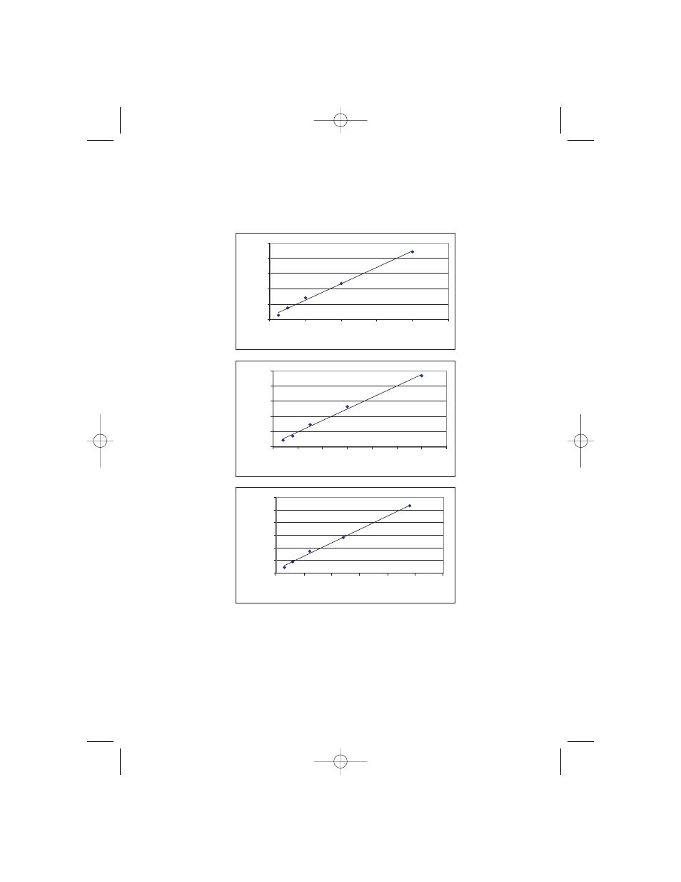
Fig. 4. Serial 2-fold dilutions of
ββ-galactosidase, run on a Criterion™ Tris-HCl 4–20% gel.
The gel was stained with Flamingo Fluorescent Gel Stain and imaged with the Molecular Imager FX
system. Fluorescence associated with the
β-galactosidase band was determined using Quantity One
®
1-D Analysis Software. A
A: Fluorescence associated with protein band in the range 0.25–4 ng. B
B:
Fluorescence associated with protein band in the range 4–60 ng. C
C: Fluorescnece associated with protein
band in the range 60–960 ng.
10003321A.qxp 8/19/2005 11:50 AM Page 9
- TransFectin™ Lipid Reagent (2 pages)
- Gene Pulser MXcell™ Electroporation System (19 pages)
- Gene Pulser MXcell™ Electroporation System (66 pages)
- Gene Pulser Xcell™ Electroporation Systems (83 pages)
- Gene Pulser® Electroporation Buffer (2 pages)
- MicroPulser™ Electroporator (31 pages)
- Helios® Gene Gun System (52 pages)
- PDS-1000 / He™ and Hepta™ Systems (51 pages)
- TGX™ FastCast™ Acrylamide Solutions (2 pages)
- Criterion™ TGX Stain-Free™ Precast Gels (16 pages)
- Image Lab™ Software (212 pages)
- Gel Doc™ EZ System (22 pages)
- Criterion Stain Free™ Tris-HCl Gels (96 pages)
- Mini-PROTEAN® TGX™ Precast Gels (52 pages)
- Image Lab™ Software (236 pages)
- ChemiDoc™ XRS+ System (42 pages)
- ChemiDoc™ XRS+ System (4 pages)
- ChemiDoc™ XRS+ System (50 pages)
- GS-800™ Calibrated Densitometer (444 pages)
- ChemiDoc™ MP System (8 pages)
- Criterion™ TGX™ Precast Gels (60 pages)
- Criterion™ Cell (13 pages)
- Image Lab™ Software (260 pages)
- Criterion™ XT Bis-Tris Precast Gels (26 pages)
- 2-D Electrophoresis Workflow (14 pages)
- Fluorescent Protein Stains (27 pages)
- 2-D Electrophoresis Workflow (22 pages)
- 2-D Electrophoresis Workflow (20 pages)
- PROTEAN® i12™ IEF System (4 pages)
- ReadyPrep™ 2-D Starter Kit (28 pages)
- EXQuest Spot Cutter (81 pages)
- Ready Gel® Zymogram Precast Gels (46 pages)
- Mini-PROTEAN 2-D Electrophoresis Cell (44 pages)
- Precision Plus Protein™ Prestained Standards (3 pages)
- Precision Plus Protein™ Unstained Standards (16 pages)
- Prestained SDS-PAGE Standards (3 pages)
- Unstained SDS-PAGE Standards (3 pages)
- Silver Stains (20 pages)
- Biotinylated Standards (3 pages)
- Biotinylated Standards (11 pages)
- IEF and 2-D Standards (3 pages)
- Mini-PROTEAN 2-D Electrophoresis Cell (20 pages)
- Mini-PROTEAN 3 Multi-Casting Chamber (10 pages)
- PROTEAN® Plus Hinged Spacer Plates and Combs (21 pages)
- Mini-PROTEAN® Tetra Handcast Systems (10 pages)