ZOLL M Series Defibrillator Rev B Non-Int 12 Lead User Manual
Page 5
Non-Interpretive 12-lead
9650-0218-01 Non-Interpretive
12-lead
-
3
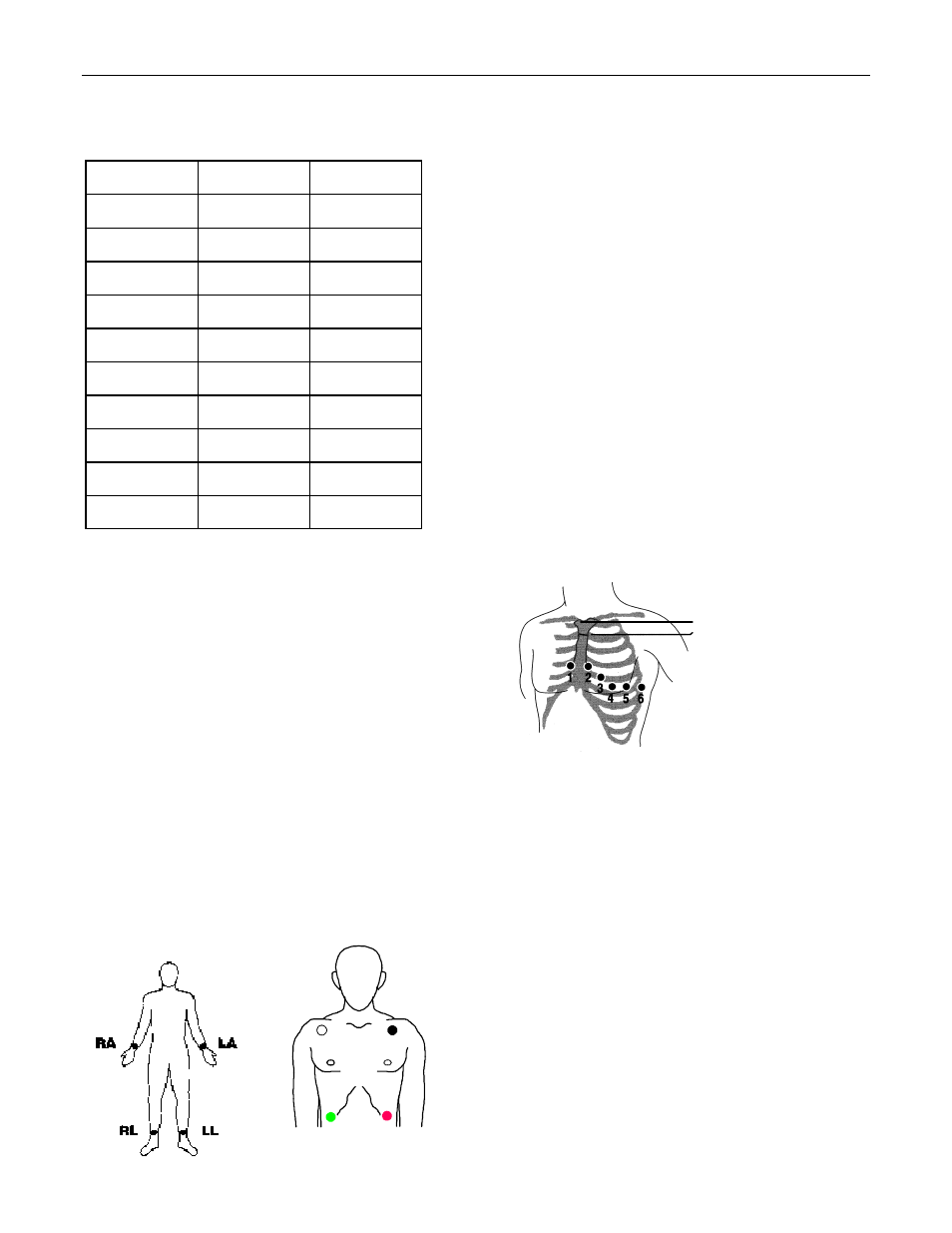
Electrode Placement
Location
AHA Labels IEC Labels
Right Arm RA (white)
R (red)
Left Arm
LA (black) L (yellow)
Right Leg RL (green)
N (black)
Left Leg
LL (red)
F (green)
Chest
V1
C1
Chest
V2
C2
Chest
V3
C3
Chest
V4
C4
Chest
V5
C5
Chest
V6
C6
Place electrodes on the patient. All electrodes must be
connected. Proper skin preparation and use of proper
electrodes are very important for a good signal quality.
If necessary prepare the patient’s skin for electrode
application by ;
• Shaving or clipping excess hair at electrode site . Avoid
placing electrodes over tendons and major muscle
masses.
• Cleaning oily skin with an alcohol pad.
• Briskly rubbing site to dry.
When acquiring 12-lead ECG from quiet supine patients,
ZOLL recommends placing the limb electrodes anywhere
along the ankles and wrists. When it is difficult for the
patient to remain motionless due to shivering, muscle
tremors, or transport vehicle movement, better results are
often obtained if limb electrodes are placed on the patient’s
thorax. (Refer to the two diagrams below for limb electrode
placement).
RA/R
LA/L
RL/N
LL/F
Place the precordial electrodes across the chest in the
following locations;
V1 : Fourth intercostal space, at the right sternal margin.
V2 : Fourth intercostal space, at the left sternal margin.
V3 : Fifth rib, between leads V2 and V4.
V4 : Fifth intercostal space, on the left midclavicular line.
V5 : Left anterior axillary line, at the horizontal level of V4.
V6 : Left midaxillary line, at the same horizontal level as V4
and V5.
Locating the V1 position (fourth intercostal space) is
critically important because it is the reference point for
locating the placement of the remaining V leads. To locate
the V1 position:
1. Place your finger on top of the jugular notch (s ee figure
below).
2. Move your finger slowly downward about 1.5 inches
(3.8 centimeters) until you feel a slight horizontal ridge
or elevation. This is the "Angle of Louis" where the
manubrium joins the body of the sternum.
3. Locate the second intercostal space on the right side,
lateral to and just below the Angle of Louis.
4. Move your finger down two more intercostal spaces to
the fourth intercostal space which is the V1 position.
Note: When placing electrodes on female patients, always
place leads V3-V6 under the breast rather than on the
breast.
Angle of Louis
Jugular notch