Measuring in scattering media, Conclusion – Ocean Optics MMS Raman User Manual
Page 25
B: Introduction to Multimodal Sampling
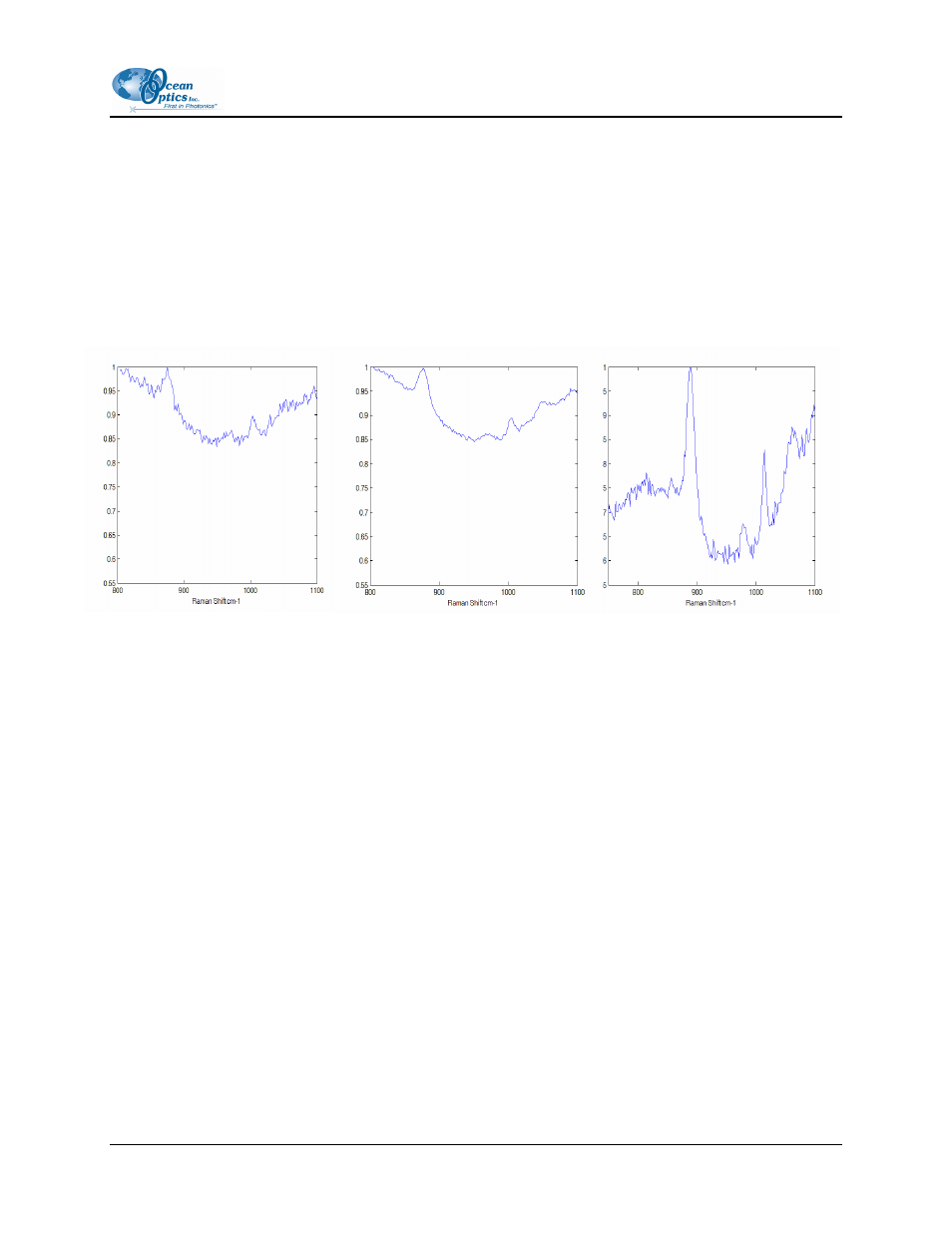
Measuring in Scattering Media
igh etendue MMS systems perform extremely well when measuring scattering samples such as blood or
ssue. For comparative purposes, we measure the Raman spectrum of ethanol in lipid. Lipid is a blood
ulating medium which offers scattering properties similar to that of blood. The Raman spectra are
own in Figures 11, 12 and 13. A higher percentage of Raman scattered photons – the signal of interest –
entering the wide area aperture of the MMS system as evidenced by the stronger primary ethanol peak
these figures. The spectra have been zoomed in to 800-1100 cm-1 range. Note that this is a 663 nm
stem, so some of the Raman ethanol peaks are lost in the background fluorescence.
Conclusion
Multimodal multiplex spectroscopy is an example of how digital instruments can be adapted to specific
measurement tasks, in this case efficient signal collection from wide area sources. Of course, sensors are
evaluated in practice by how well they perform specific tasks. For MMS systems, attractive applications
focus on molecular recognition and imaging for life and chemical science applications. While we have
shown in this white paper that MMS systems offer performance advantages over conventional systems,
the use of MMS systems as embedded biological and chemical sensors will perhaps be more significant
than their application as general purpose bench top spectrometers.
H
ti
em
sh
is
in
excitation sy
Figure 12: Raman spectrum of
ethanol in lipid with a binned slit.
Figure 11: Raman spectrum of
ethanol in lipid with pinhole
Figure 13: Raman spectrum of
ethanol in lipid
with MMS
000-40000-000-02-0906
23