Power – LW Scientific i4 12 Volt Infinity User Manual
Page 2

P. 770.270.1394
F. 770.270.2389
865 Marathon Parkway Lawrenceville GA 30046
Unpacking and Setup
Lamp Replacement
Assembly
Operation
LW Scientific packs each I-4 Microscope with utmost care. Examine the outer
and inner containers for any visual damage. Retain all of the packing material
until you have examined and tested your new microscope. If there is
damage, please contact the shipping company, as our warranty does not
cover shipping damage. If you are uncertain who the shipper is, contact the distributor from whom you purchased the
microscope. Please retain all packaging material for future use. Carefully unpack** your I-4 Microscope using the following
checklist for all the parts and accessories:
2
4
5
3
1
Remove the body of the microscope and place it on a sturdy, dust-free surface. Remove the plastic plugs in the
nosepiece. Install the objectives in such a way that when you turn the nosepiece clockwise, you are moving from the
4x, to 10x to 40x and finally the 100x objective.
Remove the microscope head from the Styrofoam carton and pull off the protective covers from the eyepiece tubes
and head mount. Insert the head mount into the upper arm of the body, taking notice that the “groove” in the flange
beneath the head must line up with a “tab” in the mounting ring on the scope (turn the head toward the side to line
up). The “groove” system is a safety feature to keep the head from falling off if the retention screw is loosened. Once
the head seats, then tighten the head retention screw to secure the head in place. Note: Do not over-tighten.
Remove plastic covers and packing material covering the stage, the condenser and lower light assembly
Insert the 10x eyepieces.
Attach the power adapter.
2
3
4
5
6
7
8
9
10
11
1
Once you have assembled all the parts and allowed your microscope to come to room temperature, plug the power
adapter into the microscope and then into the appropriate AC outlet. Note: excess cold can fog lenses and cause lamp to
fail.
Turn the light on using the black on/off switch on the rear of the scope. Next adjust the light intensity using the brightness
control wheel located on the right side of the scope.
In order to become acquainted with the controls, choose a specimen slide with which you are familiar. For example, an old
hematology slide or a commercially prepared slide. Place the slide into the slide holder by pushing back on the thumb guard
to open the slide finger. The slide finger closes slowly to eliminate the possibility of chipping the corner of your slide when it
closes.
Move the slide to the center of the stage, by turning the stage control knobs, located just below the stage. These knobs allow
you to move the slide on the X-Y axis (forward/backward and left/right).
The sub-stage iris should then be set to match the aperture of the objective for maximum resolution under each objective
power. There are numbers on the iris ring to show the correct setting for each objective power. You should begin with the 4x
or 10x objective. Only use the iris wide open when under the 100x oil objective. Closing down the iris on smaller objective
powers will improve resolution, contrast, and depth of field.
Place the filter of your choice onto the lower light assembly. Note that many customers prefer to use the blue filter for routine
use, or no filter at all.
Once you are comfortably seated, look into the oculars and move the eyepiece tubes together or apart until you see only
one complete circle of light. You have now adjusted your interpupillary distance. The I-4 binocular eyetubes can also be
rotated completely from low position to top position, which raises the eyepieces nearly 2 inches higher for tall users.
Using the 4x or 10x objectives and the coarse and fine adjustment knobs, bring the specimen into focus. Now, move the 40x
objective into place. You will feel a “clicking” action when the objective is seated properly. Again, adjust focus for best
image. You should also adjust the iris diaphragm (as described in step 5) for the best contrast and resolution.
Diopter Adjustment: Since you are using a binocular microscope, you have to adjust the normal difference in vision
between your two eyes. This is a simple but critical adjustment! Close your left eye and look into the right ocular with
your right eye. Adjust the focus to give you the best image. Now look at the ocular tube on the left. You will see that
the left ocular tube has a built-in adjustment ring. Now close your right eye and look with your left eye into the left
ocular. Using the diopter adjustment ring on the left ocular tube, adjust the focus until you see a clear, focused field.
Friction Adjustment: With repeated use and wear, the stage may drift downward out of focus. If this happens, you need only
to tighten the friction control ring (located on the right side of the microscope between the coarse adjustment and the body
of the microscope). If the coarse focus is hard to turn, you may choose to loosen the friction adjustment. There is a black
plastic friction wrench in your packaging that will engage the friction control ring to help you turn it.
Stage Stop Lever: To help prevent the stage from hitting the objectives, the I-4 Microscope is equipped with an adjustable
stage stop. Rotate the 100X oil objective into place, and put a slide into the slide holder. Slowly raise the stage, stopping when
the slide makes contact with the objective. Now, turn the stage stop lever in a clockwise direction toward you to lock the
stage from going any higher. The stage stop lever is located on the left side of the microscope between the coarse
adjustment and the body of the microscope.
Power
1 - Microscope body with Abbe condenser
2 - 10x eyepieces
1 - Binocular head (Seidentopf style)
1 - Friction adjustment wrench (for coarse focus)
2 - Filters (blue & green)
4 - Objectives 4X, 10XR, 40XR, 100XR (oil)
1 - Power adapter
1 - Warranty card
1 - Immersion oil
1 - Dust cover
**Note - Some parts may be packed in the
outer recesses of the styrofoam blocks
Ensure that the power switch is in the OFF position and that the power adapter is removed. Remove the magnetic base
condenser to expose the LED light. Unscrew the black top to remove protective glass cover and set aside. Raise the stage
as high as possible to allow better access to the LED assembly. *BE CAREFUL WHEN RAISING THE STAGE NOT TO DAMAGE
THE OBJECTIVE. Next, take a small Philip’s Head screwdriver and remove the 2 screws that secure the LED light.
Set them aside in a secure place. Using a soldering iron, gently melt the solder on the positive and negative connections
and detach the wires. Note which color wire correspnds to the positive and negative wires. Gently use a small flat head
screwdriver to remove the LED disk from the heat conductive tape. Once removed, discard the old LED light. Take the
new LED light and place it onto the adhesive tape, just as the old LED light was placed. Be sure to keep the wire
connections exposed as they will need to be connected to the new LED light.
Once the new LED light is secured to the tape and centered, solder the two wires back to the corresponding positive and
negative contact pads on the LED light. Use the two small Philip’s Head screws to secure in place. Allow 5 minutes for the
solder to set. Plug the power adapter into the back of the microscope, place the base condenser back on the
microscope, and turn the microscope on to test the LED light connection. If connection is good, screw the protective
glass cover back onto the LED assembly, and lastly, place the magnetic base condenser back into position.
If you suspect faulty electronics, call LW Scientific’s technical service department at 800-726-7345.
USE ONLY SUPPLIED POWER ADAPTER
Standard i4 Microscope:
Power Adapter Microscope:
Input: 100-240V / 50-60 Hz Auto-Switching Input: 12V 1amp
Output: 12V 1amp
Semen Evaluation Heated Stage i4 Microscope:
Power Adapter Microscope:
Input: 100-240V / 50-60 Hz Auto-Switching Input: 12V 6amp
Output: 12V 6amp
A
B
C
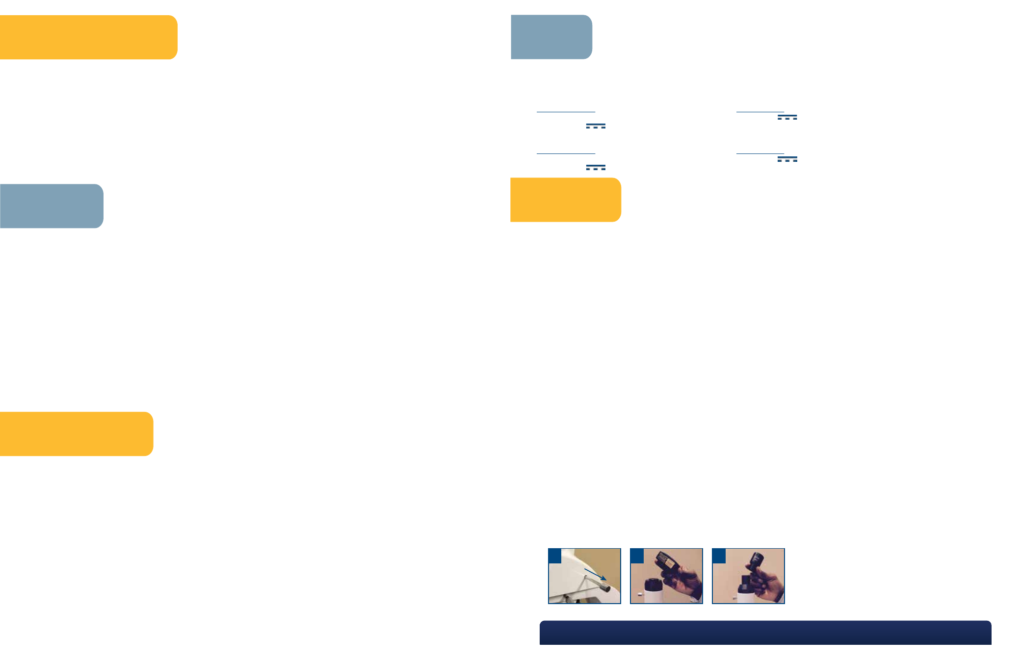
NOTE:
A: If using a trinocular head, pull out knob when
using a camera.
B: i4 Trinocular C-mount tube.
C: i4 Trinocular eyepiece tube.
