Bio-Rad Rotofor® and Mini Rotofor Cells User Manual
Page 22
heat generated during IEF keeps the temperature inside the focusing chamber
approximately 10 °C higher than that of the circulating coolant. Temperature settings
for chillers are generally between - 10°C and 4°C.
Diffusion rates of proteins are proportional to their temperature in solution.
Because proteins at steady state diffuse in and out of their focused zones it is
advisable to run the Rotofor cell at the lowest possible temperature to offset this
effect.
7.5 Electrolytes
The recommended electrolytes for the anode and cathode are 0.1 M H
3
PO
4
and 0.1 M NaOH, respectively. Because there can be a slight amount of electrolyte
exchanged through the ion exchange membranes during the focusing run, the first
one or two channels may be very acidic (
gradient in the middle channels. This will have minimal affect on the final results of
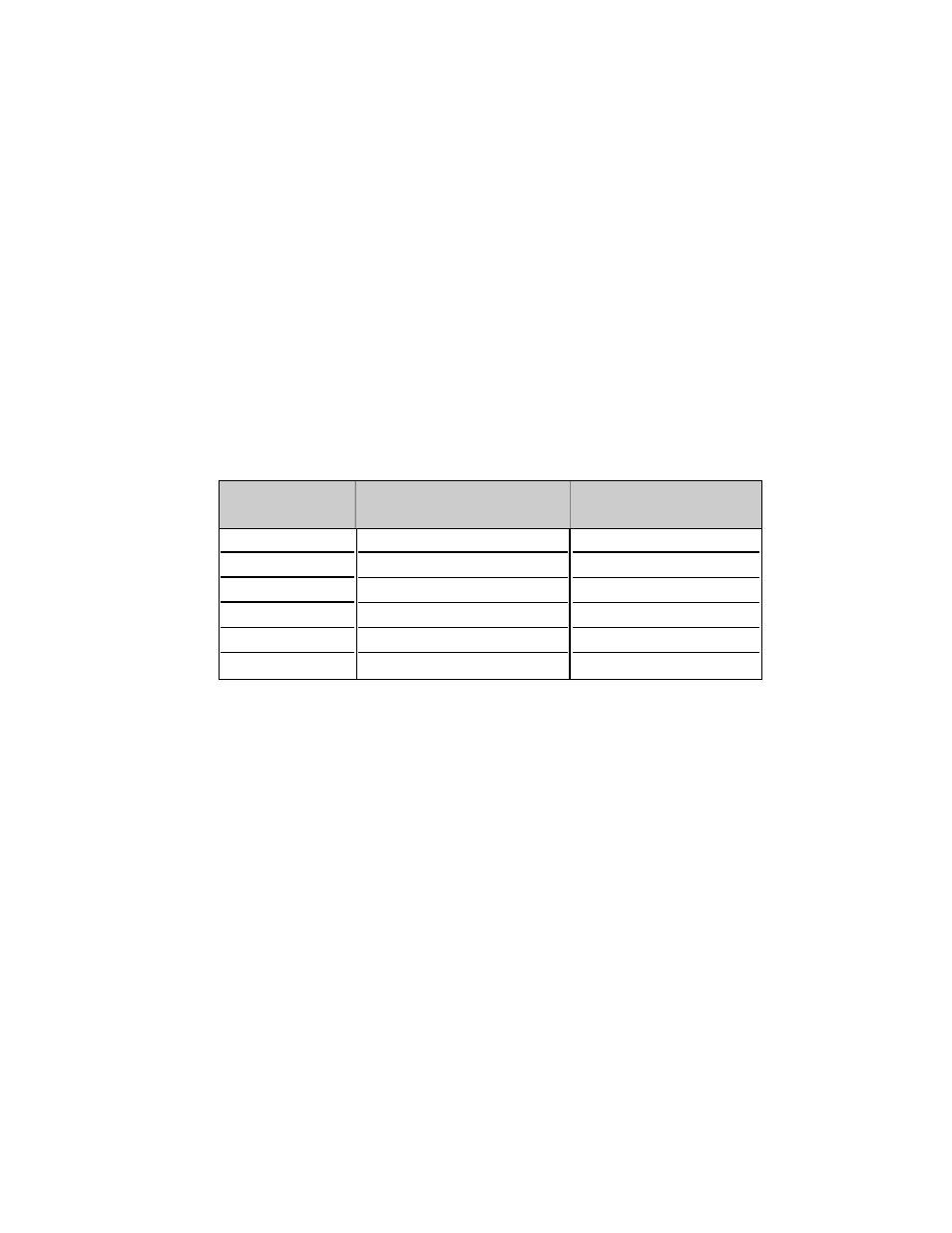
the experiment. Alternative electrolytes, e.g., amino acids, acetic acid, etc., may be
used and perform as well as H
3
PO
4
and NaOH. These include:
7.6 Pre-running the Cell
The unit should be cleaned with distilled water prior to loading the sample.
Simply fill the focusing chamber with 55 ml of distilled water and run at standard
power for 5 minutes. Drain the unit using the harvesting apparatus. This will insure
that extraneous ions have been removed from both the cell and the surface of the
ion exchange membranes.
7.7 Prefocusing
Loading the sample into the Rotofor cell is usually accomplished by injecting a
homogeneous solution of the prepared sample containing ampholytes, the protein
of interest, and any required solubility agents into the focusing chamber. However,
some proteins are especially sensitive to rapid pH shifts or to extremes of pH and
may precipitate or become denatured. To avoid exposing your protein to these
potentially damaging conditions during initial focusing, “prefocus” the focusing
media (i.e. Bio-Lyte ampholytes and solubility additives), without protein for about
an hour. This will establish the pH gradient. Then inject your protein sample into
the sample chamber at or near the point in the pH gradient that corresponds either
to the pH of the protein sample solution or the pI of your protein of interest. To
avoid disrupting the pH gradient during injection of the sample, this technique
requires that the volume of the solution containing the protein sample be as small
as possible. Prefocusing decreases exposure of proteins to rapid pH shifts and pH
extremes, minimizes the amount of time the protein spends in the Rotofor cell, and
may reduce run times by up to 50%.
18
3-5
4-6
5-7
6-8
7-9
8-10
0.5 M acetic acid
0.5 M acetic acid
0.1 M glutamic acid
0.1 M glutamic acid
0.25 M MES
0.25 M MES
0.25 M HEPES
0.5 M ethanolamine
0.5 M ethanolamine
0.1 M NaOH
0.1 M NaOH
0.1 M NaOH
pH range of
Anode
Cathode
Bio-Lyte
Electrolyte
Electrolyte