Bio-Rad ReadyPrep™ 2-D Starter Kit User Manual
Page 14
6
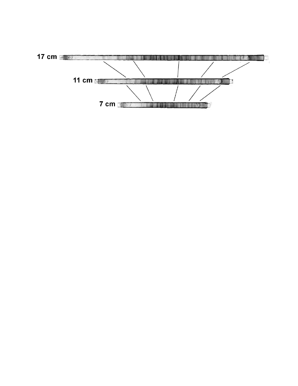
Figure 9 shows the expected pattern when IPG strips of 17 cm, 11 cm, and 7 cm are
stained with IEF gel staining solution (catalog # 161-0434) and destained with Coomassie
Brilliant Blue R-250 destaining solution (catalog # 161-0438).
4.6
Preparations Needed Before Beginning IPG Equilibration & SDS-PAGE.
Due to time considerations, it is practical to proceed to running the second-dimension
SDS-PAGE gel if either the 7 cm or 11 cm IPG strips were used. If 17 cm IPG strips were
focused, the length of the SDS-PAGE step is prohibitive in most circumstances to com-
pleting this step the same day. In this case, the IPG strips can be frozen at -70ºC as
described above (section 4.4, step 2) and the SDS-PAGE gel can be run on the next day.
1. Preparation of SDS-PAGE Gels.
a
Remove from the refrigerator the same number of 8-16% precast polyacrylamide gels
as the number of IPG strips to be run in the second dimension.
b
Open the packaging for each gel and remove the gels.
c
Remove the IPG comb from each gel and rinse the well briefly with nanopure
water using a water bottle.
d
Place the rinsed gels into a tray and when all have been processed, cover the tray
with plastic wrap to prevent the gels from drying out.
2. Preparation of 1X SDS-PAGE Gel Running Buffer.
a
Prepare sufficient 1X Tris/glycine/SDS (TGS) running buffer to run the number gels of
the size decided upon. See section 5, Appendix, for the recipe for this buffer. Bio-Rad
offers a convenient 10X TGS stock solution which is easily diluted with distilled water
to make 1X running buffer (See Section 6 for ordering information).
3. Preparation of Equilibration Buffer I and II.
The equilibration buffers should be prepared about 15 minutes before use. If the IPG strips were
frozen at -70ºC, they can be removed from the freezer and placed onto the lab bench to thaw
at this time. The strips require 10-15 minutes to thaw. It is best to not leave the thawed IPG
strips for longer than 15-20 minutes as diffusion of the proteins can result in reduced sharpness
of the protein spots.
12
Fig. 9. Isoelectric focusing of E. coli protein sample in 7 cm, 11 cm, and 17 cm IPG strips
followed by staining with IEF gel staining solution.