Luminex xMAP Antibody Coupling Kit User Manual
Page 19
Page 15 of 20
4.
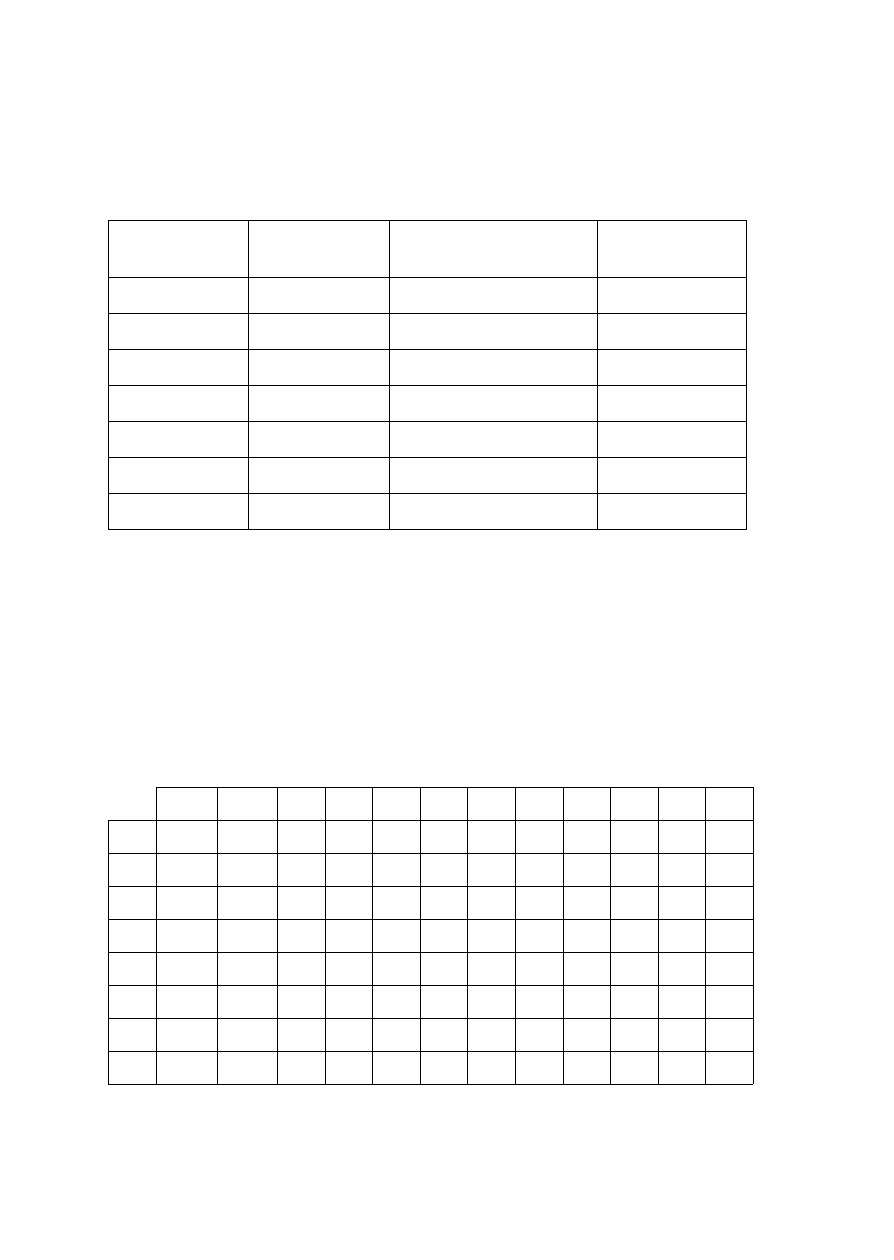
Prepare a solution of phycoerythrin-labeled anti-species IgG detection antibody at 4
μg/mL in PBS-1% BSA. Prepare a 1:2 dilution series of that detection antibody
solution to a concentration of 0.0625 μg/mL as shown in the following table.
NOTE: For optimal results use Costar round-bottom 96-well plates and Luminex
Magnetic Plate Separator with MagPlex microspheres.
5.
Aliquot 50 μL of the microsphere solution prepared in Step 3 into two entire columns
of wells of the plate (duplicate sets of 8 wells, 16 wells total).
6.
Add 50 μL of PBS-1% BSA, as a blank sample, into the wells in Row A containing
the microsphere solution.
7.
Add 50 μL of each of the diluted detection antibody solutions prepared in Step 4 into
the appropriate wells of the plate (as shown in the plate layout below).
1
2
3
4
5
6
7
8
9
10
11
12
A
Blank
Blank
B
1:64
1:64
C
1:32
1:32
D
1:16
1:16
E
1:8
1:8
F
1:4
1:4
G
1:2
1:2
H
1:1
1:1
Example plate layout using columns 1 & 2
Dilution Tube
Volume of
PBS-1% BSA
Volume of Detection
Antibody
Concentration
1:1
-
-
4 μg/mL
1:2
500 μL
500 μL from Tube 1:1
2 μg/mL
1:4
500 μL
500 μL from Tube 1:2
1 μg/mL
1:8
500 μL
500 μL from Tube 1:4
0.5 μg/mL
1:16
500 μL
500 μL from Tube 1:8
0.25 μg/mL
1:32
500 μL
500 μL from Tube 1:16
0.125 μg/mL
1:64
500 μL
500 μL from Tube 1:32
0.0625 μg/mL