Microscopic differentiation – Carolina 3M Petrifilm Yeast & Molds Count Plates User Manual
Page 7
7
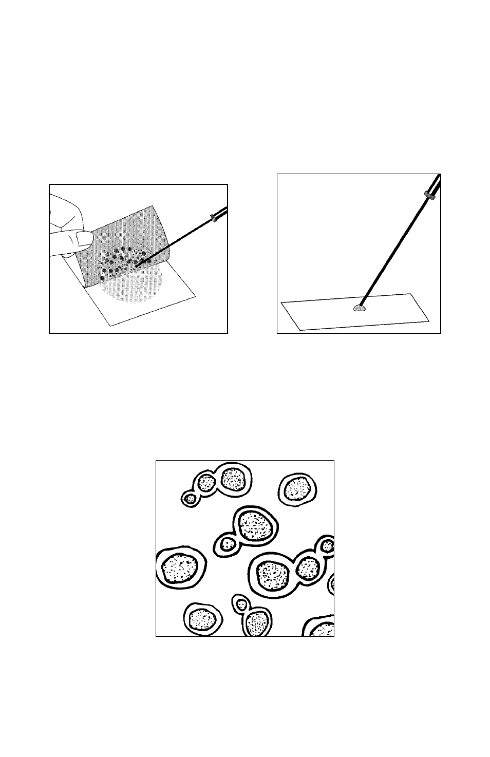
Microscopic Differentiation
To examine the organisms, lift the top film and pick the colony from the gel
(fig. 10). Transfer the colony to a drop of water on a microscope slide, cover
with a coverslip, and view under oil immersion (fig. 11). Look for oval-
shaped budding yeast (fig. 12) and for mold—both its branching mycelium
(fig. 13) and its germinating spores (fig. 14).
Figure 10
Figure 11
Figure 12
See also other documents in the category Carolina Equipment:
- NeuLog UVA (4 pages)
- NeuLog Voltage (4 pages)
- NeuLog UVB (4 pages)
- NeuLog Spirometer (4 pages)
- NeuLog Surface Temperature (4 pages)
- NeuLog Turbidity (5 pages)
- NeuLog Respiration Monitor Belt (4 pages)
- NeuLog Pressure (4 pages)
- NeuLog Relative Humidity (4 pages)
- NeuLog Magnetic (4 pages)
- NeuLog Motion (5 pages)
- NeuLog Light (4 pages)
- NeuLog Infared Thermometer (4 pages)
- NeuLog Oxygen (6 pages)
- NeuLog Photo Gate (8 pages)
- NeuLog pH (5 pages)
- NeuLog Nitrate Ion-Selective (5 pages)
- NeuLog Heart Rate & Pulse (4 pages)
- NeuLog Force Plate (4 pages)
- NeuLog Hand Dynamometer (4 pages)
- NeuLog High Temperature (4 pages)
- NeuLog Flow (4 pages)
- Corning Hot Plate Stirrer 120 V (14 pages)
- GC Series Lab Ovens (4 pages)
- 180 Series General Purpose Incubators (4 pages)
- Mold Inhibitor 876165 (1 page)
- 3M Petrifilm Aerobic Count Plates (8 pages)
- 3M Petrifilm Coliform Count Plates (12 pages)
- 3M Petrifilm Enterobacteriaceae Count Plates (12 pages)
- 3M Petrifilm E.Coli/Coliform Count Plates (12 pages)
- 44003,44004,44203,44204 Series SteamScrubber & FlaskScrubber Glassware Washers (72 pages)
- 25X Electric Pressure Steam Sterilizer (24 pages)
- NeuLog Soil Moisture (4 pages)
- NeuLog Salinity (4 pages)
- NeuLog Rotary Motion (4 pages)
- NeuLog Sound (4 pages)
- NeuLog Acceleration (5 pages)
- NeuLog Blood Pressure (4 pages)
- NeuLog Calcium Ion-Selective (5 pages)
- NeuLog Barometer (4 pages)
- NeuLog Ammonium Ion-Selective (5 pages)
- NeuLog CO2 (4 pages)
- NeuLog Conductivity (4 pages)
- NeuLog Colorimeter (5 pages)