6 determine optimum threshold – ZOLL R Series Monitor Defibrillator Rev K Operators Guide User Manual
Page 104
C
HAPTER
9
N
ONINVASIVE
T
EMPORARY
P
ACING
(O
PTIONAL
)
9–6
www.zoll.com
9650-0904-01 Rev. K
To avoid mistaking muscular response to pacing stimuli for arterial pulsations, use ONLY the
following locations for palpating pulse during pacing:
•
femoral artery
•
right brachial or radial artery
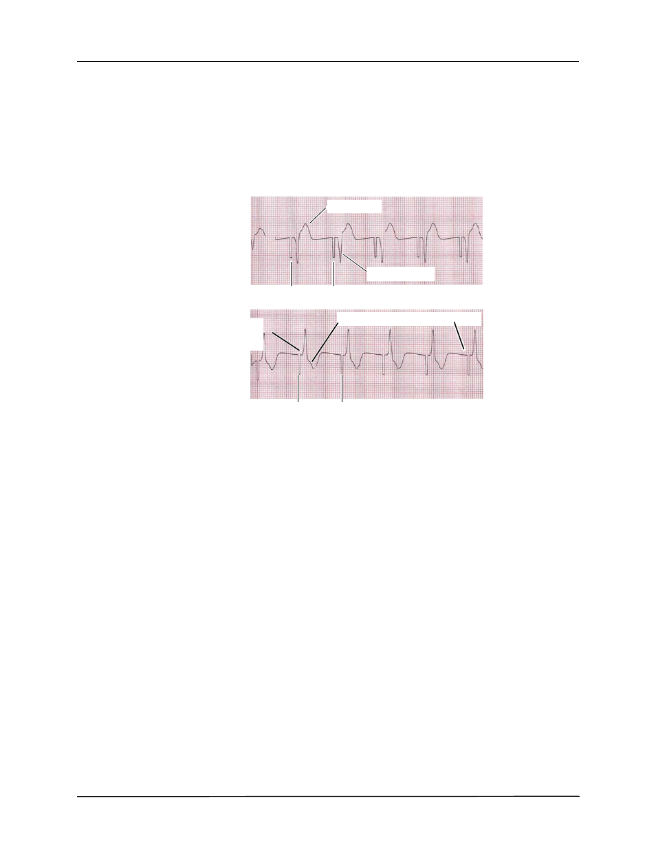
Effective pacing
The following ECG traces illustrate typical examples of effective pacing.
Changing ECG leads and size can sometimes be helpful in determining capture.
Note:
The shape and size of the paced ECG waveforms can vary depending on the ECG lead
configuration chosen; variation from patient to patient can be expected.
6 Determine Optimum Threshold
The ideal pacer current is the lowest value that maintains capture — usually about 10% above
threshold. Typical threshold currents range from 40 to 80 mA. Location of the hands-free
therapy or OneStep therapy electrodes affects the current required to obtain ventricular capture.
Typically the lowest threshold is obtained when the position of the electrodes provides the most
direct current pathway through the heart while avoiding large chest muscles. Lower stimulation
currents produce less skeletal muscle contraction and are better tolerated.
Large T wave
Negative R wave
Inverted T wave/ absence of P waves
Pacer Markers
Pacer Markers
Widened, positive QRS
(which looks like an
ectopic beat).
,