Mitosis – 3B Scientific Cell Division I Chart, Mitosis User Manual
Page 4
English
Mitosis
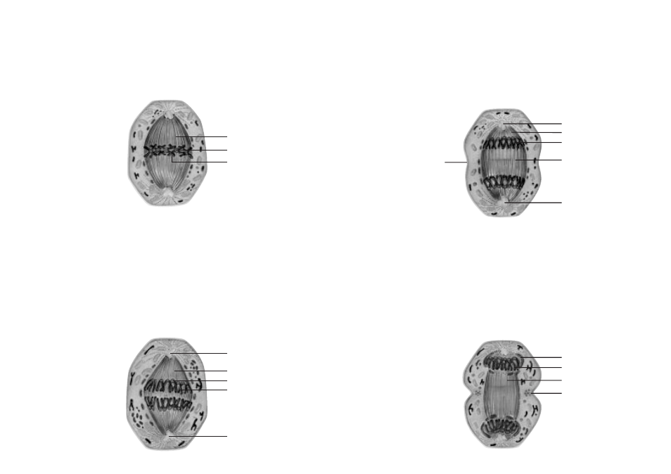
7. Later Anaphase
In the later anaphase the chromosomes (1) have reached the cell poles and now form two ”daughter”
stars. The microtubules (2) of the central spindles connected with the kinetochores at the two centrioles (3)
opposite of each other recede and disconnect. The microtubules (4) that are not connected to chromo-
somes now become longer, thus increasing the distance between the centrioles and elongating the cell.
At the equator level, the beginning stage of a cleavage furrow (5) becomes visible.
8. Telophase
In the telophase the microtubules connected with the kinetochores dissolve completely. The only remain-
ing microtubules (1) are those connecting the two cell poles with each other. A new nuclear membrane
(2) begins to form around the two separated chromosome pairs at the cell poles. The condensed DNS (3)
begins to elongate again and a new nucleolus begins to form.
The cleavage furrow at the equator level is condensed and constricts to form a ring (4), which actively
condenses the cytoplasm and leads to a further division of the new cells which are about to form.
Mitosis
English
1
1
4
5
3
1
3
2
4
3
2
4
3
2
1
4
2
3
5. Metaphase
The microtubules of the central spindle (1) have now attached precisely to the kinetochores (2) of each
doubled chromosome (3). During the metaphase the chromosomes become shorter and align exactly in
the middle between both poles of the central spindle. They form the so-called metaphase plate. Viewed
from the top, they have a star-like shape (monaster or ”mother” star).
6. Early Anaphase
In the early anaphase the previously duplicated chromatids (1) separate. In this process, the sister chro-
matids containing the same genetic information are precisely separated, forming independent chromo-
somes. This separation begins at the pairs of kinetochores (2), which is where the traction fibres of the
central spindle are attached. From here, the chromosomes are pulled slowly towards the centrioles (4)
located at the cell poles, moving along the microtubules (3) which create a traction effect as they become
shorter.