A small system that, Shows a lot of heart – GE Healthcare Vivid i User Manual
Page 4
Excellent raw data image quality. Innovative
performance features. Established clinical
tools. One-touch image optimization. The
ability to assess LV function and cardiac
performance more clearly, effectively
and confidently.
Performance features and clinical tools
• New Ultra Definition algorithms for Speckle Reduction Imaging
(SRI), Clarity and Adaptive Reject further optimize image quality.
• Smart Depth automatically adapts imaging parameters to
help save time, and increase standardization among users.
• Smart Stress helps improve workflow, shorten optimization
time and support reproducibility for review, wall segment
scoring and reporting.
• Tissue Synchronization Imaging (TSI) translates comprehensive
quantification into an easy-to-understand image demonstrating
mechanical synchronicity of different myocardial segments.
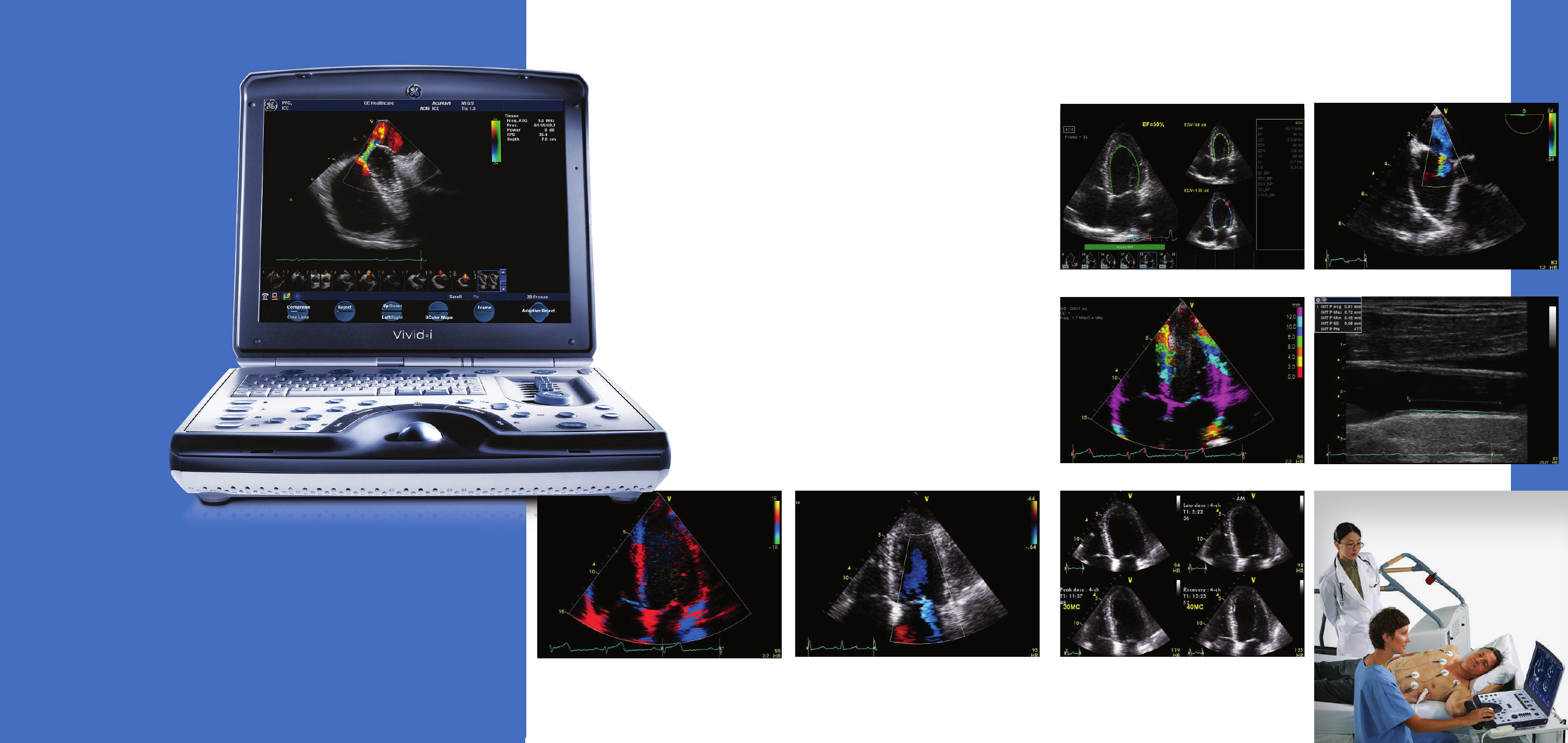
• AutoEF measurement provides the ejection fraction - one of
the most widely used clinical parameters.
Tissue Velocity Imaging apical four chamber
Mitral regurgitation apical four chamber
A small system that
shows a lot of heart.
Tissue Tracking
Common carotid artery measurement intima-media thickness
Dobutamine stress echo
Transesophageal echo with color Doppler
Auto EF