Power. performance, Heavyweight ability, Pick up-and-go portability – GE Healthcare Vivid i User Manual
Page 3: Lightweight mobility
Visualize clearly. Scan efficiently. Analyze quickly.
Vivid i’s excellent image quality and quantitative
analysis tools bring innovation to portable
ultrasound imaging.
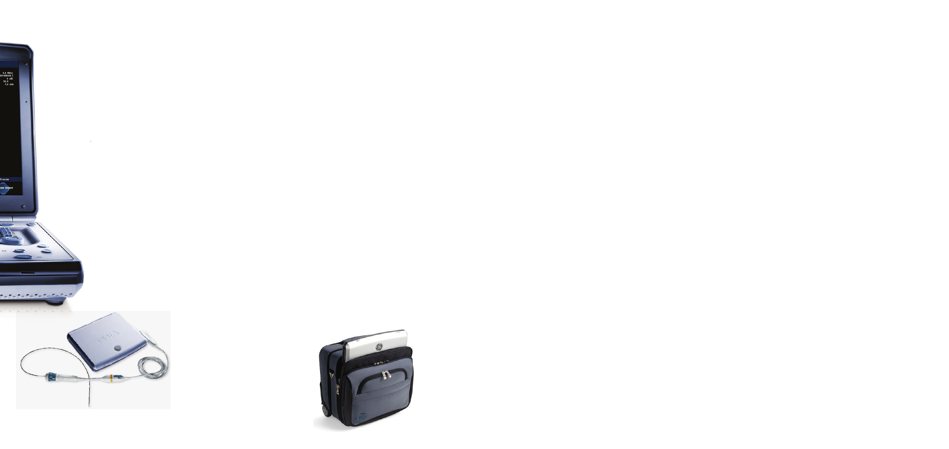
Intra-Cardiac Echo (ICE) imaging
ICE catheters deliver excellent image quality and
real-time visualization of cardiac structural anatomy,
and therapy catheters for monitoring and guidance
during interventional procedures. ICE can give you
a better understanding of structural orientation
during trans-septal puncture procedures to help
you avoid clinical complications.
Power. Performance.
Pick up-and-go portability.
OR/Anesthesia
• Supports perioperative needs with
transthoracic examinations under
challengIng conditions.
• Enable monitoring with the help of adult
or pediatric TEE.
• Support saphenous vein harvesting
and carotid evaluations.
• Use the intra-operative probe to
support specific diagnoses in the OR.
• Connect Vivid i’s TEE transducer to Vivid
console systems using an adaptor.
• Continuously scan for up to one hour
from battery.
• Share images remotely on any PC
using the eVue option, for efficient
and convenient consultations.
Obstetrics/Gynecology
• Focus on fetal echo, or comprehensive
examinations
Pediatric Echocardiography
• Examine children of all ages,
including newborns.
• Choose from a wide range of
sector, micro convex, linear and
transesophageal transducers plus
a specific ECG cable.
Shared Services
• Conduct additional vascular and
abdominal exams with Vivid i’s
comprehensive set of linear and
convex transducers.
• Display blood flow with 2D-like spatial
resolution and no color-flow-imaging
artifacts with B-Flow and BFI (Blood
Flow Imaging).
• Measuring the carotid artery’s intima-
media thickness quickly with the IMT
analysis package may obtain early
information on atherosclerosis risk.
• Wide Aperture improves the signal-to-
noise ratio and spatial resolution for
better penetration in deeper structures.
At a patient’s bedside. In the OR.
In a satellite clinic or mobile imaging
site. Vivid i’s compact size and light
weight make it easy to take excellent
ultrasound imaging performance
to any clinical environment.
With more quantitative tools and a high level
of image quality, Vivid i helps give you greater
accuracy, more diagnostic confidence and
increased productivity. All the functionality and
high performance of our full-featured premium
systems – in a portable design.
Lightweight mobility.
Heavyweight ability.
The Vivid
*
i builds on the many innovative
features and technologies of its predecessors,
incorporating new features, quantitative
analysis tools and applications that
help further improve image quality
and performance.
• Vivid i features a host of new technologies
migrated from Vivid 7 and Vivid S6, such
as the Ultra Definition image optimization
algorithms, Smart Depth, Adaptive
Reject and Wide Aperture, which provide
excellent image quality and inspire
higher clinical confidence in difficult-
to-scan patients.
• In addition to Tissue Velocity Imaging
(TVI), Tissue Tracking (TT), and Tissue
Synchronization Imaging (TSI), the
quantitative tools now include Auto EF
and on board Quantitative Analysis.
• Intra-Cardiac Echo (ICE) imaging
catheters open new application and
care areas for your ultrasound systems.
• Sixteen probes – including transthoracic
and transesophageal transducers for
cardiac adult and pediatric exams, and
linear, convex and Doppler probes –
further extend Vivid i’s wide range
of applications.
• EchoPAC
*
‘s advanced quantitative
analysis tools can be used with Vivid i’s
raw data, optimizing workflow to match
your real needs.