Protocol, Protein references – Bio-Rad Biotinylated Standards User Manual
Page 3
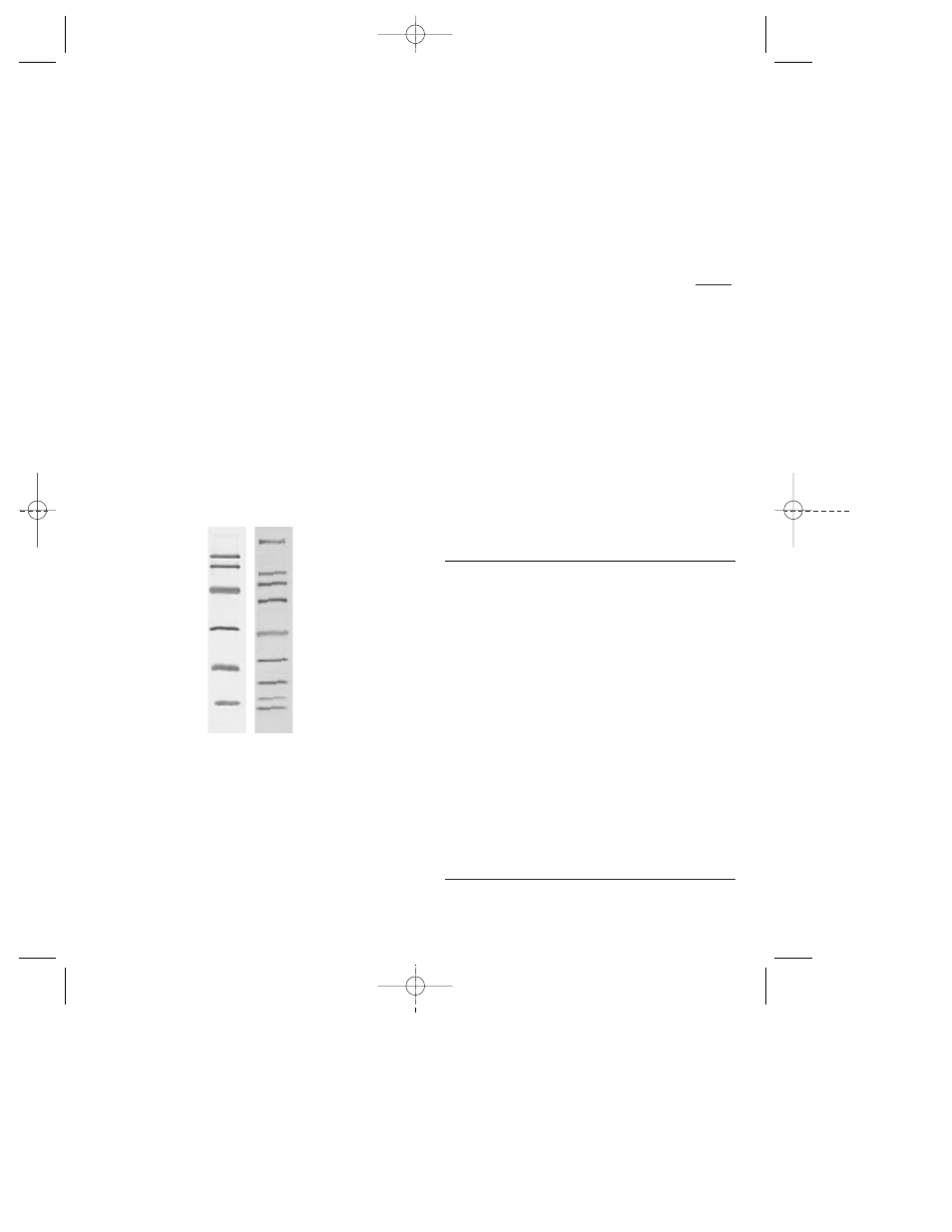
A
B
Fig. 1. Biotinylated SDS-PAGE Standards, low and
broad range. A.
Low range biotinylated SDS-PAGE stan-
dard run on a 12% gel, blotted to nitrocellulose, and
detected with Avidin-HRP.
B.
Broad range biotinylated
SDS-PAGE standards run on a 4–20% gradient gel, blot-
ted to nitrocellulose, and detected with Avidin-AP.
6
5
Protein References
Protein
Reference
Rabbit muscle myosin
Woods, E. F., Himmelfarb, S.
and Harrington, W. F.,
J. Biol.
Chem.,
238
, 2374 (1963).
E. coli
β
-galactosidase
Fowler, A. V. and Zabin, I.,
Proc.
Nat. Acad. Sci. USA,
74
, 1507
(1977).
Rabbit muscle phosphorylase b
Titani, K, et al.,
Proc. Nat. Acad
Sci. USA,
74
, 4762 (1977).
Bovine serum albumin (BSA)
Brown, J. R.,
Fed. Proc.,
34,
591
(1975).
Hen egg white ovalbumin
Warner, R. C.,
Egg Proteins
, in
The Proteins, Vol. llA, p. 435,
(Neurath, H. and Bailey, K. eds.)
Academic Press, New York
(1954).
Bovine carbonic anhydrase
Davis, R. P.,
Carbonic
Anhydrase
, in: The Enzymes,
Vol. V, p. 545 (Boyer, P. D. ed.)
Academic Press, New York
(1971).
Soybean trypsin inhibitor
Wu, Y. V. and Scheraga, H. A.,
Biochemistry,
1
, 698 (1962).
Hen egg white lysozyme
Jolles, P.,
Angew. Chem., Intl.
Edit.,
8
, 227 (1969).
Bovine pancreatic trypsin
Kassell, B. and Laskowski, M.,
inhibitor (aprotinin)
Biochem. Biophys. Res. Comm.,
20
, 463 (1965).
Phosphorylase b
Bovine serum
albumin
Ovalbumin
Carbonic anhydrase
Soybean trypsin
inhibitor
Lysozyme
Myosin
β
-galactosidase
Phosphorylase b
Bovine serum
albumin
Ovalbumin
Carbonic
anhydrase
Soybean trypsin
inhibitor
Lysozyme
Aprotinin
Protocol
1.
When using HRP conjugates, dilute standards
1:4 in sample buffer.
*
When using AP conjugates,
dilute the standards 1:20 in sample buffer. Heat for
5 minutes at 95 °C. Cool and load 10 µl/well for
mini-gels. Load 10–15 µl/well for full length gels
(16–20 cm).
2.
After electrophoretic blotting of the proteins,
detection of the biotinylated standards is per-
formed after the blocking and primary antibody
incubation steps. The avidin conjugates are used in
a 1:3,000 dilution in antibody buffer (1% gelatin in
TTBS
†
). This solution should contain the appropri-
ate dilution of blotting grade second antibody con-
jugate, protein A, or protein G conjugate. Incubate
the membrane 1 hour with gentle agitation at room
temperature.
3.
Remove the conjugate solution, and wash the
membrane twice for 5 minutes in Tris buffered
saline with 0.05% Tween-20 (TTBS)
†
with gentle
agitation. Wash twice for 5 minutes in TBS.
4.
Prepare the color development solution immediately
before use. Immerse the membrane in the solution.
Stop the development by washing the membrane in
distilled water for 10 minutes. Change the water at
least once during this time.
*
Sample buffer (SDS-PAGE reducing buffer)
Distilled water
4.0 ml
0.5 M Tris-HCl, pH 6.8
1.0 ml
Glycerol
0.8 ml
10% (w/v) SDS
1.6 ml
β
-mercaptoethanol
0.4 ml
0.1% (w/v) Bromophenol blue
0.2 ml
8.0 ml
Note: Addition of a reducing agent such as BME is
important because there is no reducing agent in the
buffer as supplied.
†
Tris buffered saline (TBS)
(20 mM Tris, 500 mM NaCl, pH 7.5)
Tris base
4.84 g
NaCl
58.44 g
Dissolve Tris and NaCl in 1.8 L distilled water. Adjust
the pH to 7.5 with HCl, and adjust the volume to 2 L
with distilled water. For TTBS add 0.5 ml Tween-20 to
1 L of TBS (0.05% Tween-20).
4
3
LIT395D 9/3/98 9:40 AM Page 3