Carolina, Overview, Safety – Carolina Mammal Heart Dissection Guide User Manual
Page 2: Procedure
Carolina
TM
Mammal Heart Dissection Guide
Overview
The Carolina Mammal Heart Dissection Guide is a general set of instructions for dissecting mammal hearts.
With each type of heart, there will be differences in the size of the structures and heart regions, but the
general structures and their relative location will be the same or very similar.
Safety
Follow safe laboratory practices when performing any dissection. Wear safety glasses or goggles, gloves, and lab
aprons when dissecting. Perform dissections on a dissecting tray or pan to contain specimens and fluids. Be
careful when using sharp instruments, such as scalpels, forceps, teasing needles, and scissors.
Procedure
1. Review the glossary provided at the end of this dissection procedure. Refer to the diagram of the heart
(on the front cover of this guide) as a general reference as you observe and identify external and internal
structures.
2. Identify the base and apex of the heart. At the base are two ear-like auricles. These are the two atria.
The rest of the heart is composed of the two ventricles. To identify the right ventricle from the left,
gently squeeze the chambers on each side of the heart. The right ventricle has thinner walls and will
compress more easily. The left ventricle has thick muscular walls due to its function of pumping blood to
the systemic circuit.
3. Cut through the wall of the right atrium and remove a portion of the wall. Be careful not to cut the right
ventricle. Observe the tricuspid valve.
4. Use a probe to push through the opening of the valve into the right ventricle. Observe the number of
flaps, or cusps, that make up this valve.
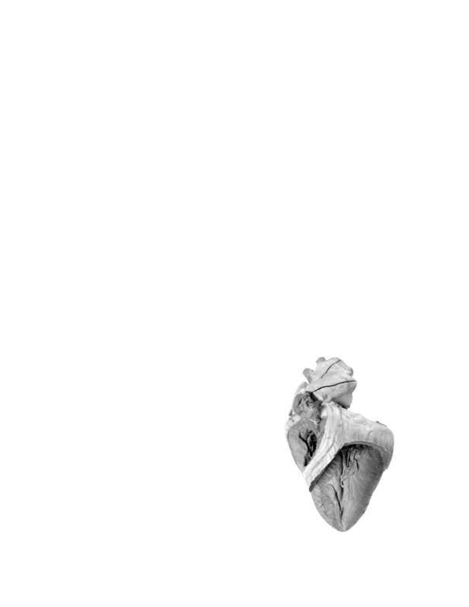
5. Refer to the dissected mammal heart image to the right.
Make an incision through the right ventricle and remove the
front portion of the wall.
6. Locate the aorta. This vessel has a larger diameter than the
pulmonary trunk and will branch immediately after leaving
the left ventricle. Cut through the wall of the aorta until you
see the aortic semilunar valve, which prevents blood from
entering the left ventricle.
7. Locate the pulmonary trunk, which is located anterior to the
aorta. Cut through the wall of this vessel until you see the
pulmonic semilunar valve, which prevents blood from
entering the right ventricle.
8. Observe the difference in the diameter of these two blood
vessels.
9. Cut through the wall of the left atrium to view the bicuspid
valve. Observe the number of cusps that make up this valve.
©2005 Carolina Biological Supply Company
Printed in USA