GE Healthcare Venue 40 User Manual
Page 4
Looking for high-resolution imaging for the needle, anatomy and vasculature? Look no further.
Because when it comes to image quality, GE’s approach to ultrasound delivers features that work
together. So, your resolution is maintained across all modes even when multiple features are
activated, including Color and Power Doppler. This also gives you deep imaging capability for larger
patients and pain management procedures. With multiple transducer options, you can perform
deep and superficial imaging – all on one system.
Superficial Imaging
Our high-frequency transducers and GE’s proprietary beamformer combine in Venue 40 to deliver
superficial anatomy in high resolution. The L8-18i-SC, our ultra-high frequency transducer, gives easier
access to superficial anatomy, in such areas as the ankle, hand and neck. Its small footprint and capability
also enable easier imaging for pediatric or smaller patients.
Needle Definition
See the target, surrounding anatomy, needle and tip defined during procedures in high resolution.
This helps you easily view the needle at various angles and depths. Our combination of
technologies can provide more accuracy and consistency to your needle-guided procedures
to help avoid complications.
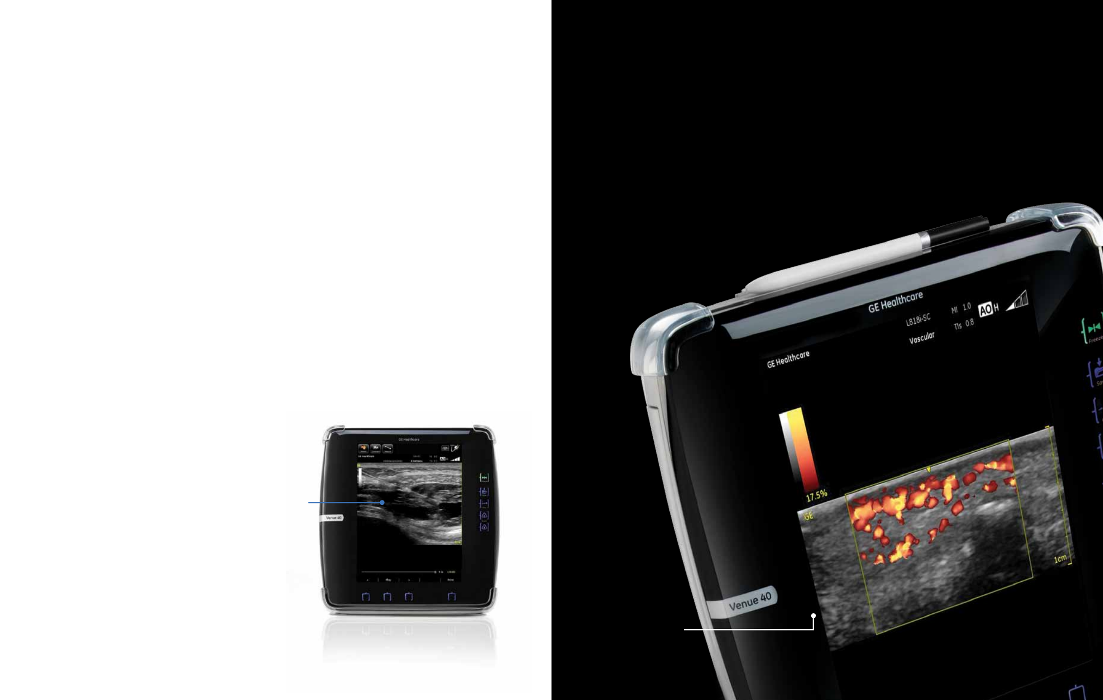
Power Doppler Imaging (PDI) sensitivity and quantification
Our proprietary beamformer detects slow blood flow in both small and large vessels.
•
PDI sensitivity helps detect small vessels, inflammation and disease states,
such as rheumatoid arthritis, tumors or clotting in both adult and pediatric patients.
•
PDI sensitivity also helps verify the presence or
absence of blood flow.
•
Color and PDI quantification helps evaluate the
amount of blood flow within a specific area, to
assist with diagnosis and monitoring.
High-resolution images
give you a higher level of confidence.
Power Doppler Imaging of the
distal digit using the L8-18i-SC
and PDI quantification.
Anesthestic surrounding Femoral Nerve.