Dedicated to bmd and beyond, Total body, body composition, Lunar dxa pediatric application – GE Healthcare DPX NT User Manual
Page 4: Bone evaluation of peripheral sites, Orthopedic - peri-prostetic hip implant, Advanced hip assessment (aha)
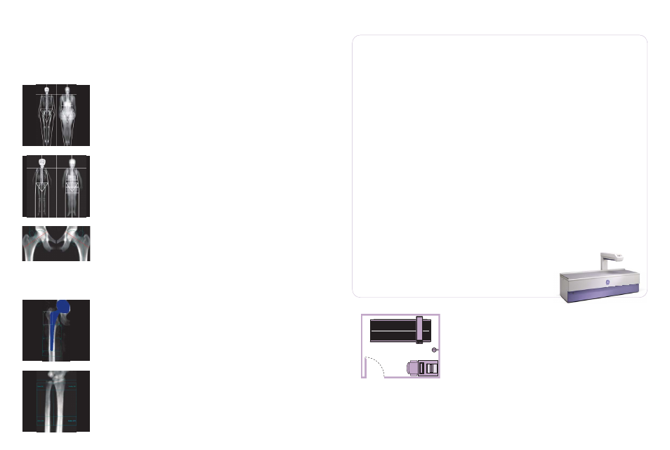
Total body, body composition
Body composition measurement with dual-energy X-ray absorptiometry (DXA) can
look beyond weight and the traditional body mass index (BMI) to determine body
fat distribution. Body composition scans with the Lunar DPX NT provide
exceptionally precise and accurate data on bone and tissue composition,
including bone mineral density (BMD), lean tissue mass, and fat tissue mass. They
provide both total body data and regional results (trunk, arms, legs, pelvis and
android/gynoid regions). The measurements are fast and non invasive.
Total body BMD - body composition
Pediatric
Orthopedic - hip implant
Forearm
Dedicated to BMD and beyond
Lunar DXA pediatric application
Now you can use one powerful set of tools to get valuable clinical information about
growth and development in children. The Lunar DXA pediatric application measures
more than BMD - it provides assessment of bone, fat and lean tissue composition.
These measurements enable enhanced evaluation of growth and development that
include height for age (bone length)
11
, BMC for bone area (bone mineralization)
11
,
bone area for height (bone width)
11
, lean body mass for height (muscle
development)
12,13
, and BMC for lean body mass (muscle-bone balance)
12,13
Bone evaluation of peripheral sites
The optional peripheral applications, such as the radius and ulna can be evaluated
to provide additional clinical information on the skeletal status of your patient, or
patient population.
Orthopedic - Peri-prostetic hip implant
The orthopedic application provides highly accurate and precise bone mineral
density and bone mineral content values. Bone assessment in the vulnerable region
surrounding an implant is now possible. This application also enables automated
bone assessment of the hip implant using standard Gruen zones (7 zones) and
extended Gruen zones (19 zones) to provide exceptional evaluation for practitioners
and clinical researchers specialized in the fields of orthopedics and surgery.
Hip Axis Length & Femur Strength Index
Advanced Hip Assessment (AHA)
The AHA application provides tools to evaluate the structural properties of the hip:
• Hip Axis Length (HAL) has been demonstrated in prospective studies as an
effective adjunct to femur bone density in predicting fracture risk.
• Cross-Sectional Moment of Inertia (CSMI) and Femur Strength Index (FSI)
are calculated for the assessment of the load-bearing capacity of the hip.
• Color bone mapping is displayed to differentiate areas of cortical and high/low
density trabecular
Lunar DPX NT technical specifications:
9,14
Available applications and options
• AP spine
• Femur
• DualFemur
• OneScan
• Advanced Hip Assessment (AHA)
• ScanCheck
• Total body/body composition
• Estimated Total Body %Fat
• Forearm
• Orthopedic
• Pediatric
• OneVision
• Composer
• Lateral spine BMD
• TeleDensitometry (e-mail)
• HIPAA SecureView
• Practice Management tools
• DICOM (worklist, color print and store)
• HL7 bidirectional interface
• Multi-User Database access (MUDB)
(1-3 or 1-10 users)
• SQL database
• Applaud CD-based training
• Remote connectivity for direct
customer support
enCORE Windows-based user interface
• Advanced intuitive graphical interface
with multimedia on-line help
• Multiple languages available
• SmartScan for scan window
optimization and dose reduction
• Automated scan mode selection
• AutoAnalysis for better precision
• Customized analysis for clinical flexibility
• Exam comparison process
• Multiple patient directories with
database
• BMD or sBMD results, BMC and area
• Extensive reference data: >12,000
USA/Northern European subjects,
as well as NHANES, and numerous
regional databases.
• T-score, Z-score, % young adult and
% age matched
• WHO guidelines for diagnosis
of osteoporosis
• Patient trending with previous
exam importation
• enCOREXpress mode for brief click path
Standard features
• Washable table pad
Quality assurance
• Automated test program with complete
mechanical and electronic tests
• Automated QA trending with
complete storage
Scanning method
• DXA pencil-beam technology
with SmartScan technology
• No scout scan required,
no moving table
X-ray characteristics
• Constant potential source at 76kV
• Dose efficient K-edge filter
• Tube current: 0.05 - 1.50 mA
Detector technology
• Nal PM tube detector
• High pulse rate
Dimensions (L x W x H) and weight
• System: 2.42m x 1.03m x 1.28m - 272kg
(95" x 41" x 50" - 599lbs)
• Table height: .63m (25")
Patient weight limit
• 136kg (300lbs)
External shielding
• Not required: X-ray safety requirements
may vary by location. Please inquire
with local regulatory authorities.
• Operating scatter: < 0.2 mR/hr (2 μSv/hr)
@ 1m (39") from X-ray source
• GE Healthcare recommends consulting
your local regulatory agency to comply
with local ordinances.
Environmental requirements
• Ambient temperature: 18-27°C (65-81°F)
• 120 VAC 50-60 Hz 20A dedicated
circuit or 230-240 VAC 50-60Hz 10A
dedicated circuit ±10%
• Humidity: 20%-80%, non-condensing
Computer workstation
• Windows platform
• Computer, printer and monitor
1. M Kamimura, H Hirabayashi, M Konishi, Q Zhou, H Kato.
Osteoporosis diagnosis and treatment decisions with Dual
Femur in Japanese women. Presented at the 17th
International Bone Densitometry Workshop, Kyoto Japan,
November 2006
2. RE Cole. Dual Femur densitometry – Effect on diagnosis and
treatment decisions. Presented at the American Society for
Bone and Mineral Research Annual meeting. October 2004.
3. RE Cole. The effect of DualFemur scanning on Osteoporosis
diagnosis and treatment decision making.
4. EN Schwartz, DM Steinberg. Performance of Computer
Assisted Densitometry (CAD) in Spine and Femur Analysis:
Comparison with Visual Assessment by Experienced
Densitometrists. Abstract Published Osteoporos Int 15
(Suppl 1):S39. Poster Presented at the IOF World Congress
on Osteoporosis, May 2004.
5. HS Barden, P Markwardt, R Payne, B Hawkins, M Frank, KG
Faulkner (2003) Automated assessment of exclusion criteria
for DXA lumbar spine scans. J Clin Densitom 6:401–409.
6. C Simonelli, L Del Rio, N Binkley. Comparison of Spine BMD
Measurements from DXA With and Without Leg Elevation.
Abstract Published J Bone Miner Res (2004) 19 (Suppl
1):S364. Poster Presented at ASBMR Annual Meeting,
October 2004.
7. M Kamimura, H Hirabayashi, M Konishi, Q Zhou, HS Barden,
H Kato. Comparison of lumbar spine BMD and T-scores
with conventional and OneScan leg positioning in a
Japanese population. Presented at the 17th International
Bone Densitometry Workshop, Kyoto Japan, November
2006
8. RH Nord, DL Ergun, KG Faulkner. Effect of patient
positioning devices on bone density measurements.
Abstract Published J Bone Miner Res (2002) 17 (Suppl 1):
S313. Poster Presented at ASBMR Annual Meeting,
September 2002.
0
9. Networking is under the user's responsibility
10. The World Health Organization (WHO), the International
Society of Clinical Densitometry (ISCD) and the National and
International Osteoporosis Foundation (NOF and IOF)
11. Molgaard C, Thomsen BL, Prentice A, Cole TJ, Michaelsen KF
(1997) Arch Dis Child 76:9-15.
12. Crabtree NJ, Kibirge MS, Fordham JN, Banks LM, Muntoni F,
Chinn D, Boivin CM, Shaw NJ (2004) The relationship between
lean body mass and bone mineral content in paediatric
health and disease. Bone 35:965-972.
13. Schoenau E, Neu CM, Beck B, Manz F, Rauch F (2002) Bone
mineral content per muscle cross-sectional area as an index
of the functional muscle-bone unit. J Bone Miner Res 17:1095-
1101.
14. Depending on product configuration and availability.
Contact GE Healthcare or our local distributor for the
detailed current configuration and optional hardware.
8' (2.4m)
10' (3.0m)
Minimum room dimensions:
References