Ne w, Cube, 3d asl – GE Healthcare Signa HDxt 1.5T User Manual
Page 9: High-density head-neck-spine array, Bravo
Cube
*
Cube replaces several plane-after-plane 2D
acquisitions with a single 3D volume scan —
providing you with T2, T2 FLAIR, or PD contrast.
Easily reformat sub-millimeter isotropic volume
data from a single acquisition into any plane
without gaps — and with the same resolution as
the original plane.
ARC is an innovative, auto-calibrating, data-
driven, parallel imaging method designed to
reduce scan time and streamline reconstruction
with high accuracy. In addition, it enables small
fields of view during prescription.
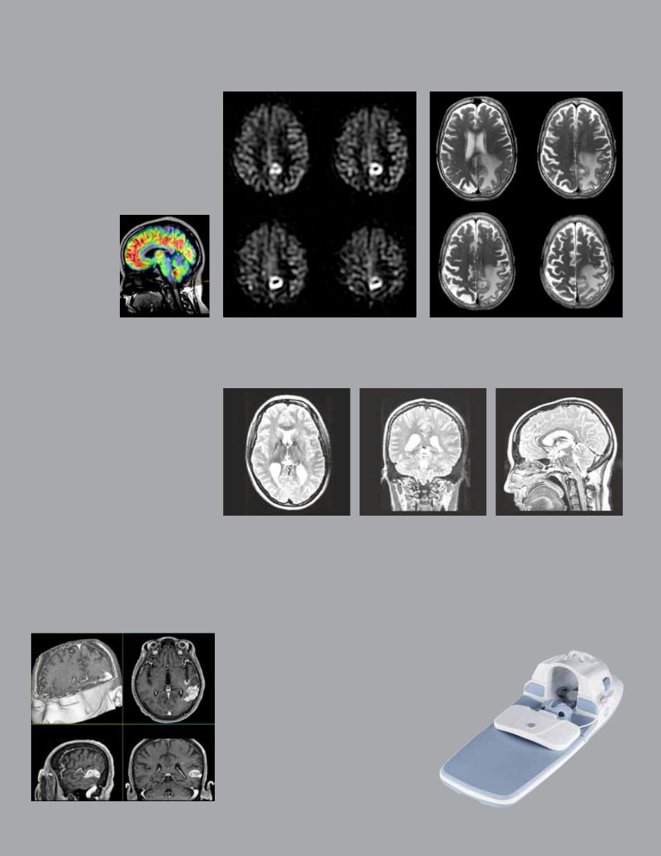
3D ASL
3D ASL is a robust non-contrast tissue perfusion
imaging technique that provides quantitative
assessment of cerebral blood flow (CBF)
and color perfusion maps (bottom). The
hyperintense signal in the left parietal occipital
lobe on this axial T2 image (far right) correlates
well with clearly depicted hyperperfusion on
the 3D ASL image (right). Note excellent SNR
throughout the entire anatomy.
High-Density
Head-Neck-Spine Array
Image the head, neck, and spine
without changing arrays or
repositioning the patient.
And, do it with high definition.
BRAVO
A 3D inversion-prepared
SPGR technique, BRAVO
provides high resolution
T1-weighted brain images
with optimized grey-white
matter contrast, using GE’s
innovative self-calibrating
ARC parallel imaging
technique. This 3D image
of a patient with brain
metastasis was acquired
with 1 mm
3
voxel.
NE
W
GE Healthcare Signa HDxt 1.5T Optima Edition
9