Capture images with clarity – GE Healthcare LOGIQ P6 User Manual
Page 4
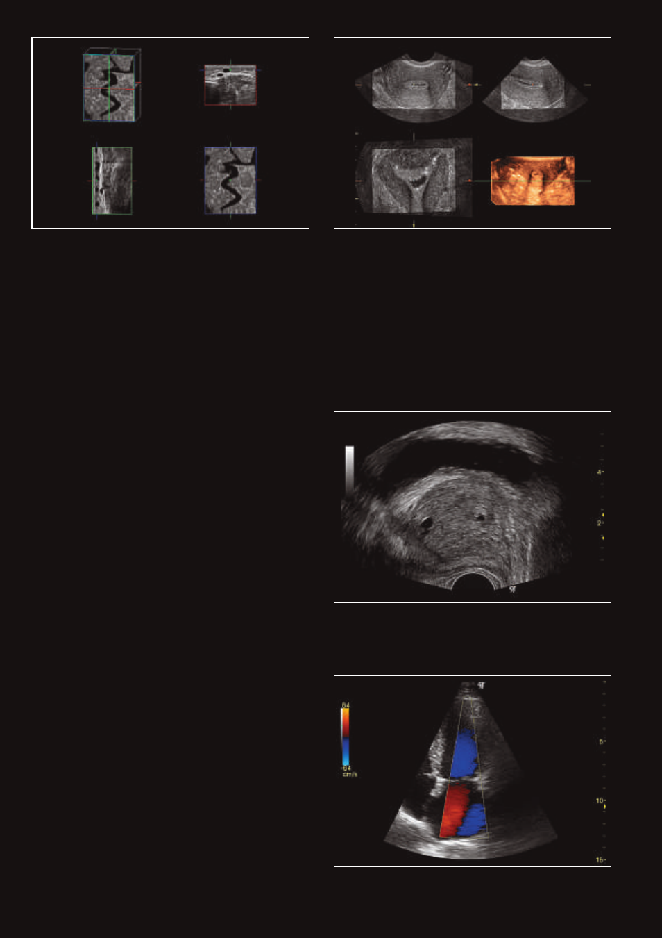
Varicose vein displayed using Easy 3D; additional clinical
information about the C-plane and the course of the vessel
can be collected while acquiring a 3D image
3D volume acquisition of the uterus using the 4DE7C probe
during a Saline Infusion Sonography (SIS) procedure
Sagittal prostate showing cystic structure imaging with the
E8CS probes using fundamental imaging and SRI
Apical four-chamber view of adult heart with color Doppler
using the 3S probe
Capture images with clarity.
It’s simple. A clear image leads to the most
confident decision. That’s why LOGIQ P6
is designed using innovative leadership
technologies from GE Healthcare. Starting
with an enhanced beamformer for deeper
penetration, higher resolution and better color
flow sensitivity, you’ll find these advancements
in imaging and more:
Speckle Reduction Imaging (SRI). Heighten your visibility of
organs and lesions with high-definition contrast resolution
that suppresses speckle artifact while maintaining true tissue
architecture.
CrossXBeam
™
. Enhance tissue and border differentiation with
a real-time spatial compounding acquisition and processing.
Real-time 4D Imaging. Acquire and construct volumetric
images, displaying multiplaner views of the anatomy. See
anatomical relationships not visualized otherwise.
DualBeam. Maintain high frame rates while using high
line density, even in abnormal settings. Increase temporal
resolution in fast-flow cardiac and vascular studies.
Harmonics. Increase resolution and cystic clarity with a
combination of coded harmonics and Phase Inversion
Harmonics.