Glossary – Carolina Mammal Eye Dissection Guide User Manual
Page 3
10. Place the cornea on your dissecting tray and cut it in half to observe its thickness.
11. Insert the forceps through the opening created and carefully separate the edge of the iris from the inner
surface of the eye. You may be able to
remove the iris intact.
12. Pick up the back half of the eyeball and
observe the structures on the inside. Identify
the retina, which contains the cone and rod
receptor cells. Follow this mass of nerve cells
to their convergence at the back of the eye,
where the optic nerve begins. This is called
the blind spot.
13. Turn the back half of the eyeball over and
observe the optic nerve on the outside of the
eye. Pinch the nerve with your forceps to see
the separate fibers of the nerve.
14. Look at the interior of the back portion of
the eyeball again. Move the retina so that
you can see the dark, metallic-looking tissue
at the back of the eye. This is the choroid, a thin layer that lies between the retina and the sclera. The
portion of the choroid that appears iridescent blue and green with shades of yellow is called the tapetum.
15. Once you have observed all the structures of the eye, dispose of the specimen in accordance with local
guidelines and your teacher’s instructions.
Glossary
Aqueous humor - clear fluid filling the area between the lens and the cornea, composed mostly of water;
helps maintain the shape of the eyeball.
Blind spot - area of the retina where the receptor cells converge to form the optic nerve.
Choroid - thin, dark sheet of tissue between the retina and the sclera.
Cones - receptor cells of the retina that are responsible for perceiving color.
Cornea - transparent covering that allows light to enter the eye; on a preserved specimen, the cornea is cloudy.
Hyaloid fossa - indention in the center of the vitreous body that supports the lens.
Iris - diaphragm that regulates the size of the pupil.
Lens - biconvex transparent structure that focuses the light coming in through the cornea and pupil.
Optic nerve - bundle of nerve cells that send signals from the eye to the brain.
Pupil - opening through which light enters the eye.
Retina - light-sensitive portion of the eye composed of receptor cells called cones and rods.
Rods - receptor cells of the retina that are responsible for perceiving difference in light intensity.
Sclera - outer covering of the eyeball; a tough, opaque sheet of connective tissue that protects inner
structures of the eyeball and helps maintain rigidity.
Tapetum - iridescent portion of the choroid tissue.
Vitreous body - the cavity between the retina and the back of the lens.
Vitreous humor - viscous fluid that fills the vitreous body; helps maintain the shape of the eyeball.
Zonula ciliaris - ligaments that suspend the lens and stretch it to focus vision.
T e a c h e r ’ s M a n u a l
3
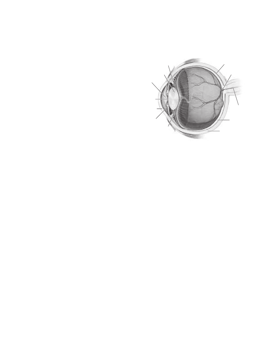
Sclera
Retina
Optic nerve
Blind spot
Choroid
Zonula ciliaris
Lens
Aqueous humor
Pupil
Cornea
Iris
Hyaloid fossa
Vitreous
humor