Carolina, Overview, Safety – Carolina Mammal Kidney Dissection Guide User Manual
Page 2: Procedure
Carolina
TM
Mammal Kidney Dissection Guide
Overview
The Carolina Mammal Kidney Dissection Guide is a general set of instructions for dissecting mammal
kidneys. With each type of kidney, there will be differences in the size of the structures and kidney regions,
but the general structures and their relative location will be the same or very similar.
Safety
Follow safe laboratory practices when performing any dissection. Wear safety glasses or goggles, gloves, and lab
aprons when dissecting. Perform dissections on a dissecting tray or pan to contain specimens and fluids. Be
careful when using sharp instruments, such as scalpels, forceps, teasing needles, and scissors.
Procedure
1. Review the glossary provided at the end of this dissection guide. Refer to the diagram of the kidney as
a general reference as you observe and identify external and internal structures.
2. Observe the renal capsule. This structure is made up of dense, irregular connective tissue and provides
protection as well as helps maintain shape. Remove any adipose tissue that may be attached to the
capsule.
3. Locate the hilus. This is an indentation where the ureter and blood vessels enter and exit the kidney.
Remove excess adipose tissue to observe the ureter more closely. The renal artery and vein may be
difficult to locate; they were severed close to the hilus when the kidney was removed from the animal.
4. Make a frontal section through the kidney.
Locate the cortex and medulla. The
medulla lies below the cortex. Observe
and record the appearance of each region.
5. The medulla consists of numerous conical
structures called renal pyramids. The base
of each pyramid lies next to the cortex,
while the tip forms a renal papilla. Each
papilla projects into the renal sinus.
Locate the renal pyramids, renal papilla,
and renal sinus.
6. Renal pyramids are separated by bands of
tissue called renal columns. Each column
begins in the cortex and extends through
the medulla. Examine the texture of this
tissue. Columns have a granular texture
similar to that of the cortex.
7. Each renal pyramid and adjacent cortical
region make up a renal lobe. Urine
production occurs in the renal lobes. Each
renal papilla discharges urine into a cup-shaped minor calyx. Four or five minor calyces merge to form a
major calyx. Major calyces merge to form the renal pelvis. Using a probe, trace the path of urine from the
renal pyramids to the renal pelvis.
©2005 Carolina Biological Supply Company
Printed in USA
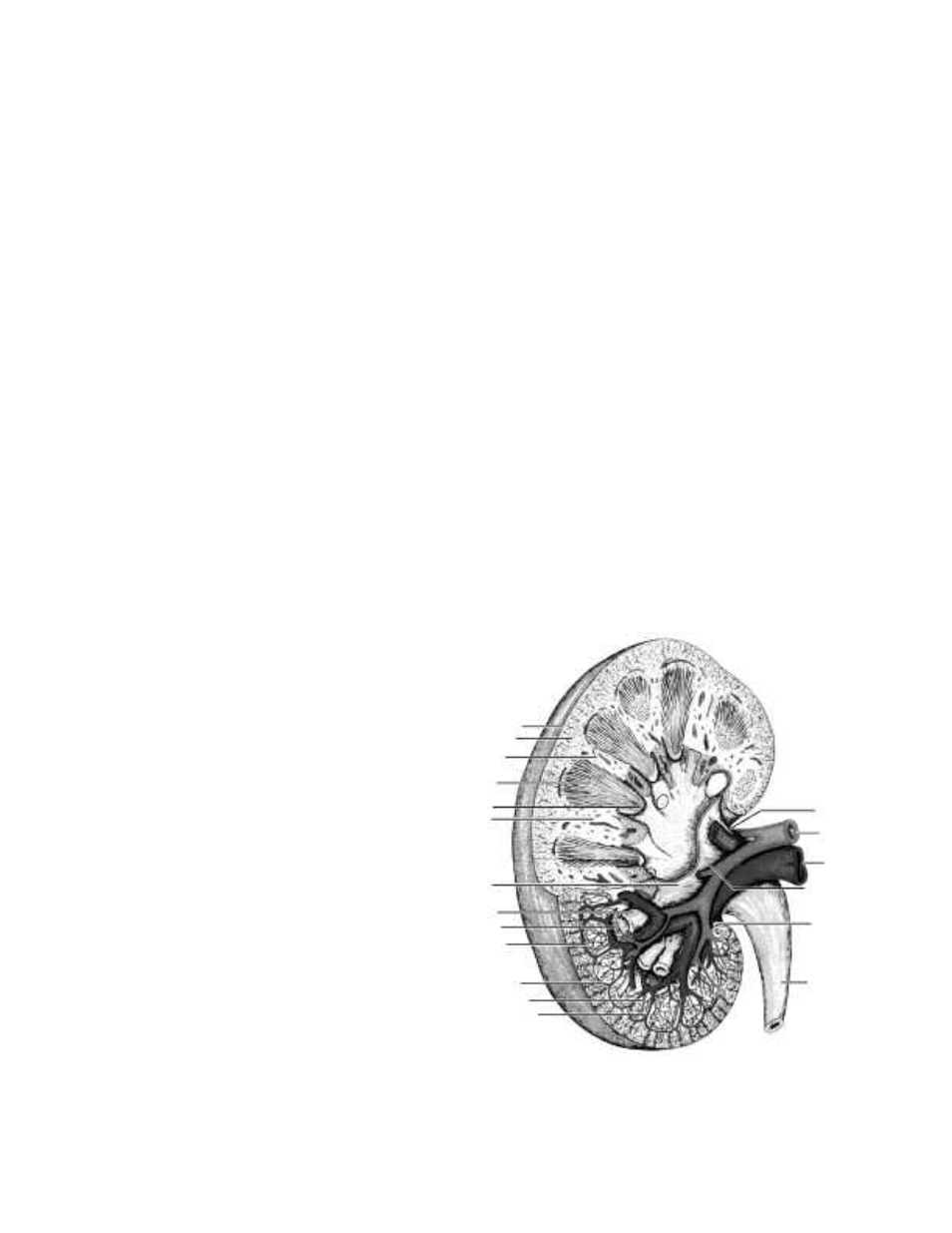
Capsule
Cortex
Medulla
Pyramid
Papilla
Column
Major calyx
Minor calyx
Pyramid
Arcuate artery
Arcuate vein
Interlobar artery
Interlobar vein
Ureter
Sinus
Pelvis
Renal vein
Renal artery
Hilus