Fluke Biomedical 84-317 User Manual
Page 6
Nuclear Associates 84-317 & 84-317-7000
Operators Manual
1-2
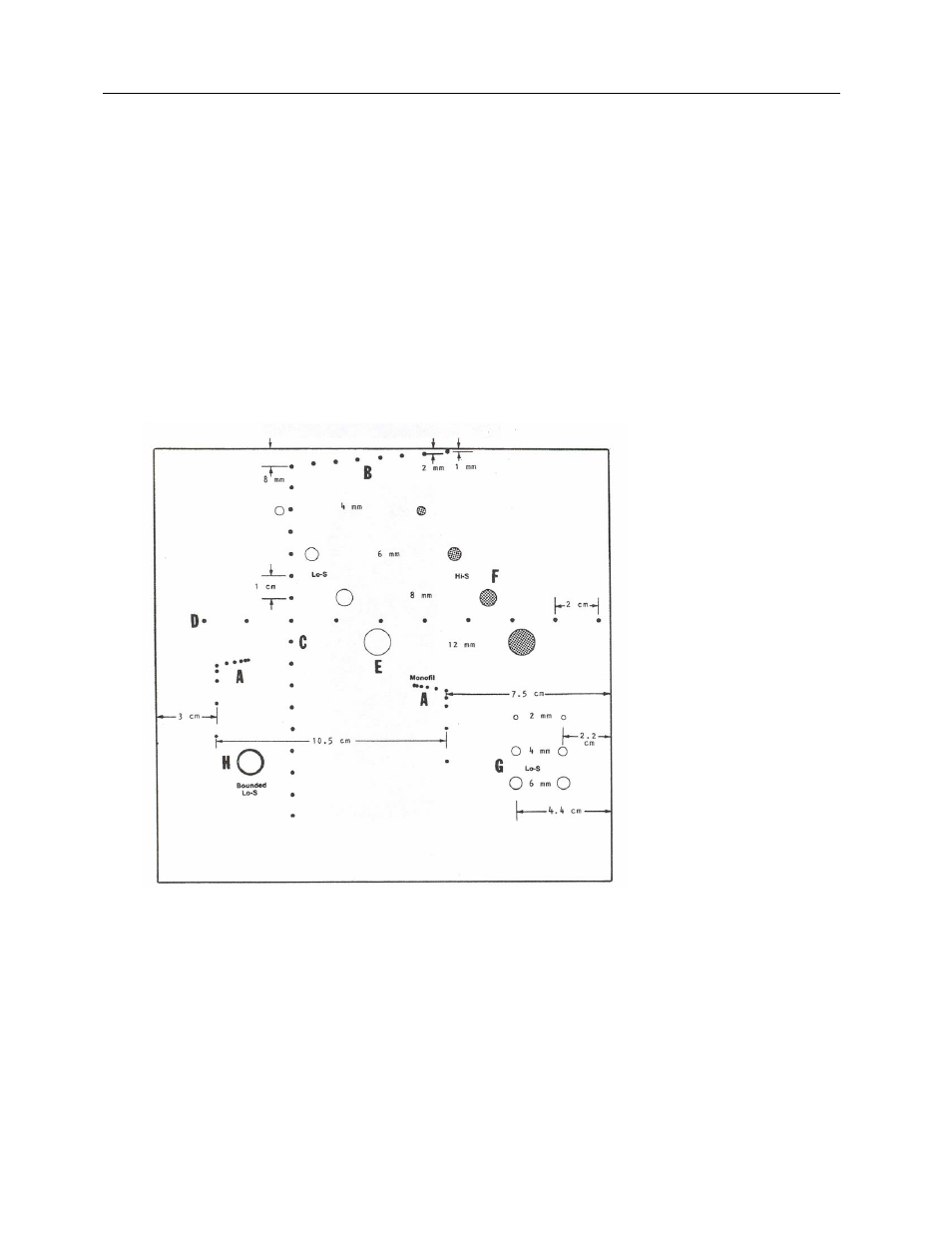
E. A diagonal row of four (Lo-S) simulated cysts (E, Figure 1-1) has cylinders of gel similar to the
background filling material of the phantom. The "cysts" (4, 6, 8 and 12 mm D.) have no scattering
centers (hence "Low-scatter" or "Lo-S") and are "transparent' to the ultrasound beam.
F. Another row of four cysts (F, Figure 1-1), parallel to the first and of the same size, is filled with gel
having more scattering centers than the background material. It is labeled "Hi-S" (high scatter) and
simulates solid lesions similar to tumors.
G. Six more "Lo-S" cysts are in two columns in the low right corner (G, Figure 1-1). Each column
contains a 2 mm, 4 mm and 6 mm cyst. One column is at 2.2 cm and the other at 4.4 cm from the
side-scanning surface. These are used with short-focal-Iength transducers and scanners designed
for close viewing.
H. One cyst-like object (Bounded Lo-S) (H, Figure 1-1) is encased in a highly reflecting plastic skin. It
represents normally fluid-filled organs such as the larger blood vessels.
I.
The filling medium is a hydrogel containing a scattering agent that, at 72°F, simulates human liver
parenchyma with respect to attenuation, scattering and propagation velocity over the range of
frequencies used in ultrasonic scanning. Sealed into a sturdy plastic case, it requires little or no
care. The scanning surfaces are made of soft plastic film that simulates skin.
Figure 1-1.
Internal Targets