Fluke Biomedical 18-250 User Manual
Page 6
Nuclear Associates 18-250
Operators Manual
1-2
1.2 Accreditation Images on Recumbent or Upright Digital
Stereo Biopsy Units
1. Center the phantom at the chest wall edge of the image receptor. Place the compression paddle in
contact with the phantom to secure it.
2. If phototiming is available, make a phototimed exposure.
3. If the unit does not have phototiming, manually select the kVp and mAs routinely used to
radiograph the Mammographic Accreditation Phantom (Model 18-220).
4. Make
the
exposure.
5. Analyze the image on the monitor.
6. To pass accreditation with the Digital Mammography Evaluation Phantom, you must be able to see
3 fibers,
3 groups of specks, and 2.5 masses.
7. Record your image on disk or film.
8. In accordance with the American College of Radiology, this test should be done weekly.
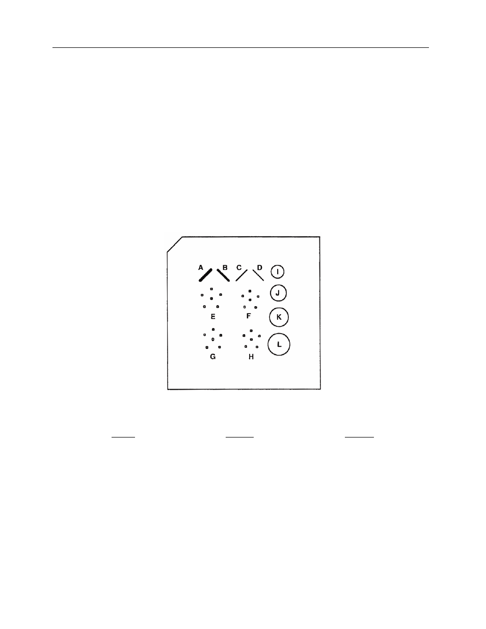
Wax Insert
Fibers Specks Masses
A. 0.93 mm nylon fiber
E. 0.54 mm speck
I. 0.25 mm (thickness) mass
B. 0.74 mm nylon fiber
F. 0.32 mm speck
J. 0.50 mm (thickness) mass
C. 0.54 mm nylon fiber
G. 0.24 mm speck
K.0.75 mm (thickness) mass
D. 0.32 mm nylon fiber
H. 0.20 mm speck
L. 1.0 mm (thickness) mass