Section, Overview sample volumes pre-concentration dilution, Sample requirements/preparation – Iris Sample Processing StatSpin® CytoFuge 12 Versatile Cytocentrifuge System User Manual
Page 11
6
Section
3
Sample Requirements/Preparation
Overview
The approximate cell concentration of the specimen should be established prior to slide preparation on
the Cytofuge 12. Samples containing higher than optimal cell concentration will produce slides with cells
too closely packed or overlapping. Samples containing too low a cell concentration will yield slides where
cells are difficult to find, count, or examine. The following is a general guideline for sample concentration
based on an average cell diameter of 10 µm:
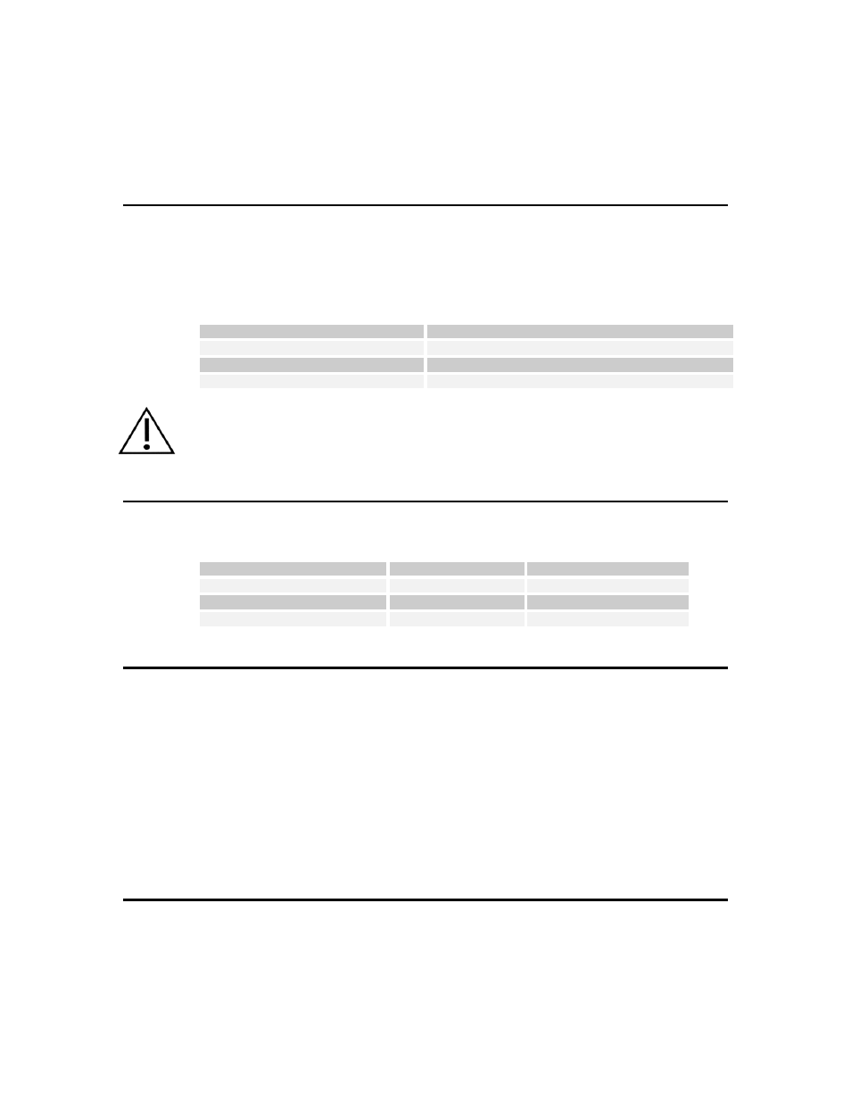
Sample Concentration
Recommendation
500 - 1500 cells/µL*
Use sample as is
< 500 cells/µL
Pre-concentrate sample
> 1500 cells/µL
Dilute sample
* In samples known for low cell populations (e.g. CSF), 50-100 cell/µL will produce acceptable results.
CAUTION
–
Follow Universal Precautions with all biological specimens, regardless of whether a
specimen is known to contain an infectious agent. Bioseals cannot be relied upon as a sole
safeguard against contamination by pathogens. Always load and unload the rotor under a biological
safety hood (See references).
Sample Volumes
Use the following guidelines for sample volumes in each of the Cytofuge concentrators:
Max. Volume Range
Optimum Volume Range
Filter Concentrator
50 to 300 µL
100 to 300 µL
One-well Cell Concentrator
300 to 1600 µL
400 to 800 µL
Three-well Cell Concentrator
50 to 400 µL
100 to 200 µL
Pre-concentration
For best results, samples low in cellular content should be pre-concentrated. For example, if the original
sample contains about 100 cells/µL, a 10x pre-concentration will provide the 1,000 cells/µL, which is
recommended for the Cytofuge 12.
To pre-concentrate a sample,
1.
Transfer 10 mL of sample to a conical polypropylene centrifuge tube.
2.
Spin the sample at 1000-1500 xg for 10-15 minutes in a conventional centrifuge.
3.
Decant 9 mL of cell-free supernatant.
4.
Mix the cell pellet and remaining supernatant by vortexing or agitation of the tube.
5.
Pipette appropriate volume of the concentrated sample to Cytofuge concentrators.
Dilution
For best results, dilute samples that have an extremely high cell density. For most applications, buffered
saline or standard tissue culture media (e.g. Geys balanced salt solution) may be used as a diluent.
Adding a drop or two of bovine serum albumin (BSA) promotes cell adhesion to the microscope slide.